the MOUSE
FUOTY 2022 6th Place. Old Legend
- Joined
- Sep 27, 2023
- Posts
- 1,991
- Reputation
- 4,164
Abstract
Testicular torsion (TT) is a common cause of acute scrotal pain in young men and accounts for as many as 26% of cases of acute scrotum. Accurate diagnosis and differentiation of TT from acute epididymo-orchitis is essential because TT is treated surgically and epididymitis with or without orchitis is treated medically. Distinguishing TT from epididymo-orchitis is generally a clinical dilemma. Sonography remains the best imaging modality for these entities, but the sonographic findings can be complex and can mimic complicated epididymo-orchitis when dealing with partial TT and torsion-detorsion syndrome. This case report illustrates the complex sonography findings of surgically proven partial TT with torsion-detorsion syndrome.Introduction
Testicular torsion (TT) is a true urologic emergency and must be differentiated from other causes of acute testicular pain because a delay in diagnosis and management can lead to loss of the testis and infertility.1–5 Testicular torsion can occur at any age but commonly affects 1 in 4000 males younger than 25 years old each year.1,4 Diagnosis of partial testicular torsion (PTT) and torsion-detorsion syndrome (TDS) is challenging as changes in the testis depend on the duration of torsion and the degree of rotation of the spermatic cord. Torsion-detorsion syndrome or intermittent testicular torsion (ITT) is defined as acute and intermittent sharp testicular pain due to impeded blood flow, interspersed with asymptomatic intervals. Sonography remains the best imaging modality to evaluate for acute scrotal pain.1,6–8 However, there is overlap in the gray-scale and Doppler findings of epididymo-orchitis (EO) and PTT with TDS, and testicular or paratesticular hyperemia during the detorsion phase of TDS can be confused with complicated EO.5,8,9 The clinical presentation of TT and acute EO also is often similar. Diagnosis of ITT is important as these patients are at high risk to develop acute torsion. Intermittent spermatic cord torsion can be treated with elective testicular fixation. Misdiagnosis or delay in diagnosis may create acute unresolved torsion and potential testicular loss.10Case Report
A 43-year-old man presented to the emergency department due to sudden onset of sharp left testicular pain. The patient’s history was significant for recent EO, which was treated with antibiotics 2 weeks prior. At this visit, he was given intravenous (IV) Ciprofloxacin for a clinical diagnosis of EO; the patient remained stable without sharp pain and was discharged home with pain medication.The patient returned to the emergency department a few hours later with uncontrolled left scrotal pain, low grade fever, and leukocytosis with a white blood cell count of 17.7. On physical examination, his left testis was swollen, erythematous, and tender to palpation. A clinical diagnosis of EO was again made, however, scrotal sonography was ordered to exclude TT. Scrotal sonography was performed utilizing a Philips iU-22 ultrasound system (Philips Medical Systems; Bothell, WA) with a 12-5 MHz linear transducer. Color Doppler imaging and spectral Doppler velocity measurements were done in addition to gray-scale imaging. Gray-scale images showed the testes to be symmetric, with normal echogenicity (Figure 1). Color Doppler imaging showed normal blood flow in the right testis (Figure 2), slightly increased blood flow in the left testis (Figure 3), and diffusely increased blood flow in the left epididymis (Figure 4). Spectral Doppler evaluation showed a normal low resistance arterial flow waveform in the right testis (Figure 5). Areas of absent (Figure 6) and reversed (Figure 7) diastolic flow were noted within the left testis. Differential diagnosis based on the sonographic features was severe complicated EO versus partial and intermittent testicular torsion.


The patient was admitted to the urology service and started on IV antibiotics. Urine and blood cultures were negative. A few hours later, physical examination showed that the left testis had become larger and extremely tender, with associated scrotal edema, and the patient’s symptoms continued to worsen despite IV antibiotics. A repeat sonogram was performed 4 hours later to re-evaluate the left testis. Echogenicity of the left testis remained normal and isoechoic to the right testis (Figure 8). Color and spectral Doppler examination again showed normal blood flow in the right testis with a normal low resistance arterial flow waveform (Figures 9 and 10). Decreased blood flow was seen in the left testis (Figures 11 and 12); additional findings of note were increased left scrotal wall thickening with associated hyperemia (Figure 12) and a left hydrocele (Figure 13). Decreased diastolic flow was seen in the left testis; however, diastolic flow reversal in the left testis was no longer appreciated (Figure 13). Venous flow within the left testis was present (Figure 14).


A diagnosis of PTT and TDS was made based on the clinical and sonographic findings, and the patient was immediately taken to surgery. At surgery, the left testis was dusky with poor blood flow and a left orchiectomy was done. A right orchiopexy was performed prophylactically to prevent torsion. Histopathology of the left orchiectomy specimen showed tubules with normal spermatogenesis, marked interstitial edema and hemorrhage, and early necrosis of the interstitial cells of Leydig consistent with torsion (Figure 15).

Discussion
The most important objective in patients with sharp, acute scrotal pain is to exclude or confirm TT. The differential diagnosis of the acutely painful scrotum includes TT, trauma, epididymitis/orchitis, incarcerated hernia, and torsion of the appendix testis.9 Accurate clinical distinction between TT and EO is difficult in up to 50% of cases and is generally a clinical dilemma. The differentiation between these two entities is crucial because TT is treated surgically and epididymitis with or without orchitis is treated medically. Testicular torsion requires urgent intervention to avoid organ loss and infertility. The optimal time frame for testicular salvage is less than 6 hours after the onset of symptoms.5,9Testicular Torsion
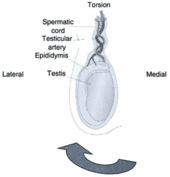
The annual incidence of TT is 1 in 4000 males younger than 25 years.1–4 Torsion of the spermatic cord usually occurs in the absence of any precipitating event. Only 4% to 8% of cases are a result of trauma. Other factors predisposing patients to TT include an increase in testicular volume (often associated with puberty), testicular tumor, testis with a horizontal lie, a history of cryptorchidism, and a spermatic cord with a long intrascrotal portion.2,3,5Torsion of the spermatic cord initially obstructs venous return. Subsequent equalization of venous and arterial pressures compromises arterial flow, resulting in testicular ischemia. The degree of ischemia and resultant changes in the testis depend on the duration of torsion and the degree of rotation of the spermatic cord. Ischemia can occur as soon as 4 hours after torsion and is almost certain after 24 hours.3,4
Ipsilateral absent cremasteric reflex is the most accurate sign of TT on clinical examination.11 This reflex is a contraction of the cremaster muscle in response to lightly stroking the inner upper thigh (via the sensory and motor fibers of the genitofemoral nerve); this muscle contraction elevates or pulls up the testis on the side stroked. On physical examination, horizontal lie of the affected testis with the patient in a standing position may also suggest the diagnosis of ITT.2
Sonography
The sensitivity of color flow Doppler to TT in pediatric patients is 90% to 100%, and specificity is nearly 100%. In the adult population, a similar sensitivity of 80% to 98% and specificity of 97% to 100% have been reported.1,12 The sensitivity of spectral Doppler ranges from 67% to 100%. An understanding of normal blood flow to the testes is important in interpreting color Doppler and spectral Doppler waveforms in the setting of TT. The normal spectral waveform of the testicular artery has low-resistance flow. Resistive indices (RIs) in normal intratesticular arteries range from 0.48 to 0.75 with a mean of 0.62.13Gray-scale and Doppler findings in TT include the following:1,6,8
•
Normal echogenicity of the testis in the early phase.
•
Hypoechoic testis after 4 to 6 hours secondary to edema.
•
Heterogeneous testis after 24 hours secondary to hemorrhage and infarction.
•
Reactive hydrocele and skin thickening.
•
Enlarged twisted spermatic cord superior to the symptomatic testis—“whirlpool sign.”
•
Decreased or absent flow in symptomatic testis.
•
Normal or decreased flow in testis with incomplete torsion (<360 degrees).
•
Normal or increased flow in testis and paratesticular soft tissues from reactive hyperemia with detorsion.
•
Absence of a dicrotic notch resulting in a monophasic waveform on spectral Doppler.
•
Increased resistance to arterial flow with a decrease in diastolic flow velocity on spectral Doppler.
•
Reversal of diastolic flow on spectral Doppler.14,15
In the small case series by Cassar and colleagues, most of the cases of PTT were diagnosed only after careful examination of the morphologic and spectral Doppler waveform characteristics as compared to the contralateral testis or a different region within the same testis.6
Identification of a sonographic “whirlpool sign” related to twisting of the spermatic cord is reported to be the most specific and sensitive sign of both complete and incomplete torsion and may help to make a definite diagnosis.16,17 The whirlpool sign is an acute rotation of the spermatic cord observed while scanning its transverse plane in which the spermatic cord looks twisted or spiraled, similar to a whirlpool.
Treatment
Treatment of TT involves rapid restoration of blood flow to the affected testis. The optimal time frame is less than 6 hours after the onset of symptoms. In one study, investigators quoted a testicular salvage rate of 90% if detorsion occurred less than 6 hours from the onset of symptoms; this rate fell to 50% after 12 hours and to less than 10% after 24 hours.18 Manual detorsion by external rotation of the testis can be successful, but restoration of blood flow must be confirmed following the maneuver. Surgical exploration provides definitive treatment for the affected testis by orchiopexy and allows for prophylactic orchiopexy of the contralateral testis.Epididymitis and Epididymo-Orchitis
Epididymitis and epididymo-orchitis are also common causes of acute scrotal pain in adolescent boys and adults.9 In adolescents, many instances are secondary to sexually transmitted organisms such as Chlamydia trachomatis and Neisseria gonorrhea. In prepubertal boys and in men older than 35 years of age, the disease is most frequently caused by Escherichia coli and Proteus mirabilis. The epididymis is the organ primarily involved in EO, with orchitis developing in 20% to 40% of cases due to direct spread of infection.Early in the course of the disease, on physical examination the epididymis can be palpated as an enlarged tender structure separate from the testis. Scrotal pain associated with epididymitis is usually relieved when the testes are elevated over the symphysis pubis (the Prehn sign). This sign may help clinically differentiate between epididymitis and torsion of the spermatic cord, in which scrotal pain is not lessened with this maneuver.
Sonography
The sensitivity of color Doppler imaging in detecting scrotal inflammation is nearly 100%. In 20% of cases of epididymitis and 40% of cases of orchitis, hyperemia is seen with color Doppler even when gray-scale findings are normal.7,9Gray-scale and Doppler findings include the following:
•
Enlarged hypoechoic or hyperechoic epididymis (presumably secondary to hemorrhage).
•
Reactive hydrocele or pyocele with scrotal wall thickening.
•
Testicular enlargement and a heterogeneous testicular echotexture.
•
Hyperemia of the epididymis and testis with color Doppler.
•
Decreased vascular resistance on spectral Doppler.
•
Reversal of flow during diastole, suggestive of venous infarction.19
Increased RIs and reversed diastolic flow have been reported in PTT, and also as a complication with severe EO.6,17,19 In severe EO, reversal of diastolic flow is caused by swelling and edema occluding the venous outflow and implies the risk of impending infarction.


