



Askinov

𝖋𝖚𝖙𝖚𝖗𝖊 𝟔'𝟏 𝖍𝖙𝖓 𝖘𝖑𝖆𝖞𝖊𝖗
- Joined
- Oct 31, 2025
- Posts
- 4,381
- Reputation
- 10,166


So we all sigma right ?? sigma boys never go and do the real surgery instead we do it ourselves that's why here is my cheap knock off of lateral commissuroplasty
he he
if you are from my old you it looks like ts
Do not open this spoiler if you are from my old thread
why not view more videos ??
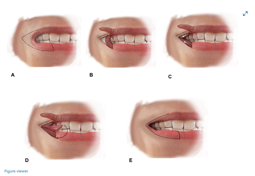
although this is a lift that le op wanted to add but the surgical process is identical so you can take a rough idea
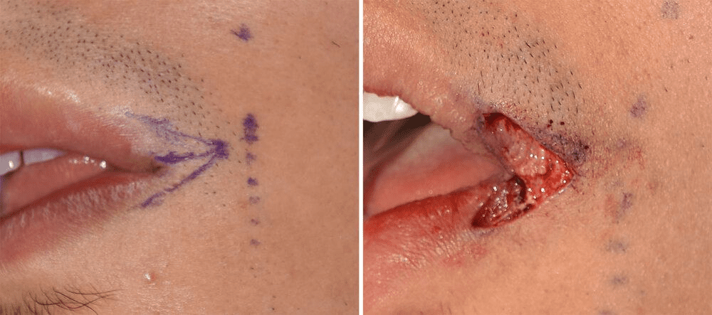
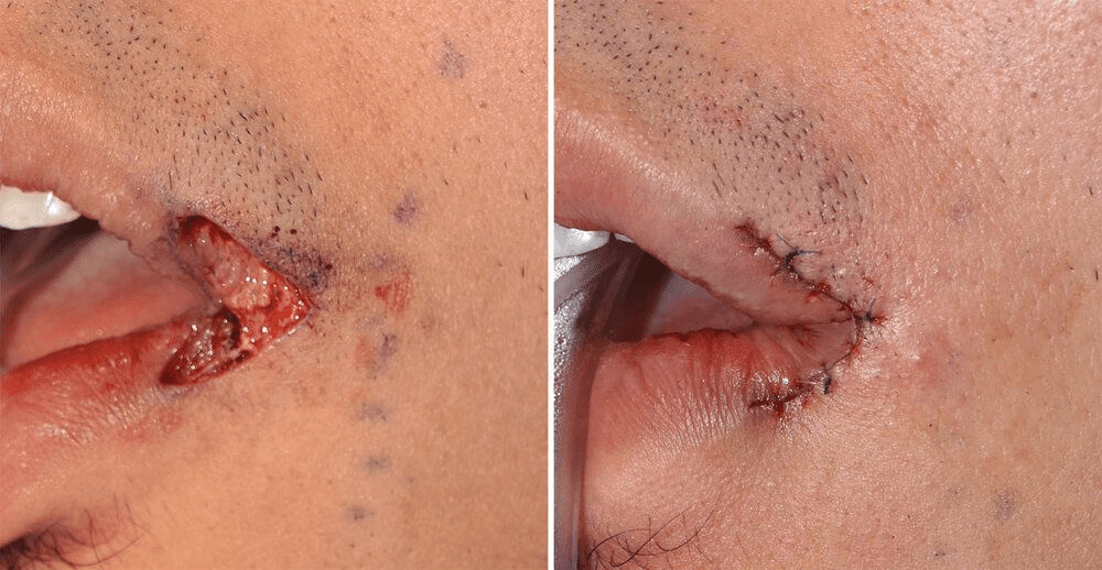
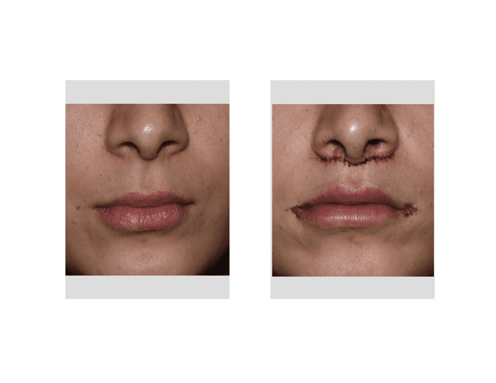
as always since the op is lazy he gonna copy paste how this procedure is done from his old guide
The surgeon identifies the desired new width for the mouth. A common aesthetic landmark is the mid-pupillary line (a vertical line dropped from the center of the pupil).
A horizontal incision is made starting from the existing corner of the mouth and extending outward to the new marked position.
Tissue Excision: Small triangular segments of skin are removed above and below the horizontal incision line. This creates a "gap" or defect that allows the corner of the mouth to be moved outward.
Muscle Adjustment: A small wedge of the orbicularis oris muscle (the muscle surrounding the lips) is often excised to create space for the new corner and prevent the mouth from "relapsing" to its original smaller size.
The inner lining of the mouth (buccal mucosa) is mobilized and pulled forward (advanced) to cover the newly created gap. This creates the new pink "vermilion" part of the lip in the widened area.
Layered Closure: The surgeon uses fine, permanent, or dissolvable sutures to close the wound in multiple layers—internal mucosa, muscle, and external skin—to ensure strength and minimize scarring
So bhai you know our skin is divided into various section ?? .It consist of
1st layer epi dermis
2nd dermis
3rd hypodermis
so water right
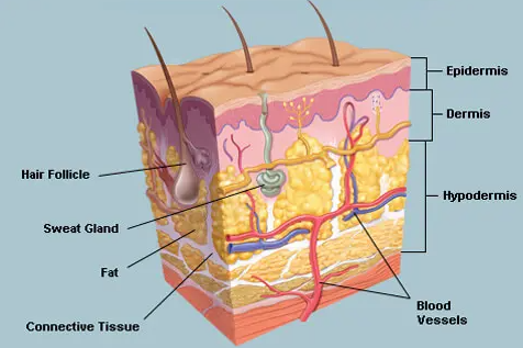
you know where does the scar actually becomes a problem ?
Regeneration of Dermis: Scarring and Cells Involved - PMC
There are many studies on certain skin cell specifications and their contribution to wound healing. In this review, we provide an overview of dermal cell heterogeneity and their participation in skin repair, scar formation, and in the composition of ...
 pmc.ncbi.nlm.nih.gov
jfl at this muh research paper just google it bhai

and so yeah don't cut after dermis just go up to middle of dermis you must be fine
+ you don't wanna go deep cause there are veins and tissue if damage occurs in them it can lead to permanent scarring and will look bad ofc
the problem is you never know which blade to choose so you can go up to middle dermis dwbi chad askinov is here to help ya
1st thing you need to keep on mind is that there is no blade that can cut only up to dermis to cut only up to dermis we can get precision blades that can control up to dermis see not cut up to dermis only but with these blade you can control up to dermis
imo surgical blades are op in this situation
eg
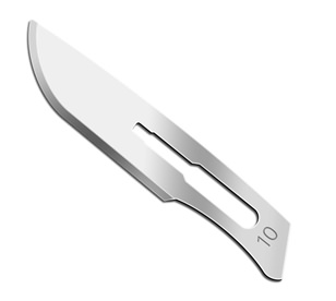
1. #10 scalpel blade
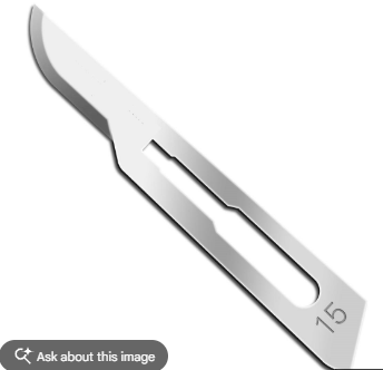
2.#15 scalpel blade
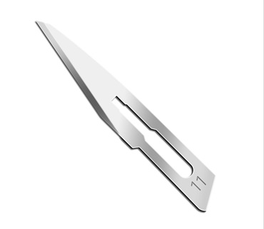
3.#11 scalpel blade
4.Dermatome Blades
5.Humby/Cobbett Knife Blades
Trust me you are not using the last two blades
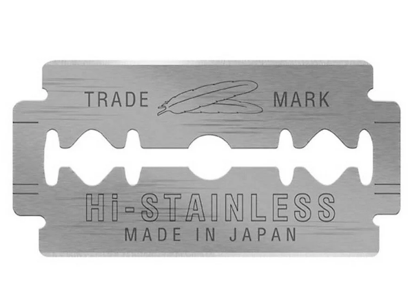
instead you also can use a sharp kitchen kife lightly with almost to no control in depth and you can also use your grandpa blade shaving blade ig but you also can not control the depth in here use it very lightly and don't use rusty one ofc like sayori told
Facial Antiseptics
There are few option available eh we use antiseptics to ensure the site you are cutting at is free of bacteria ( water )
so no future risk of infection can occur i have few examples of Antiseptics which can be used at face
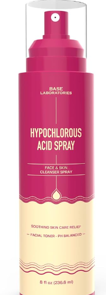
1. Hypochlorous acid ( 0.005% to 0.02% )
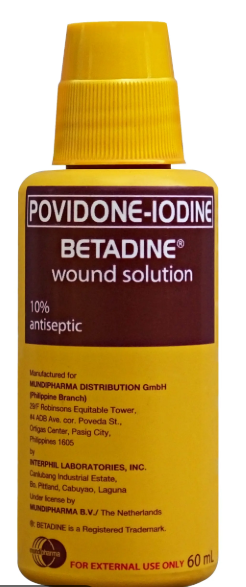
2.Diluted povidone-iodine ( 0.35% to 1% )
3.dettol for our indian brahs
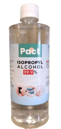
4.Isopropyl (rubbing) alcohol
5.Hydrogen peroxide ( 1% to 3% )
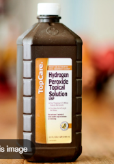
Problem with some antiseptics is that some burns like h202 and rubbing alcohol so after cutting you can get high level of irritation and pain while cutting so i will say to avoid these
how to apply antiseptics then ??
1st wash your face with warm water
2nd let it dry
3rd use a cotton pad to apply these just apply a bit use this cotton pad
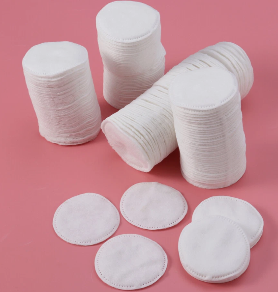
these can also be used to soak up excess blood
apply with these
There are few option available eh we use antiseptics to ensure the site you are cutting at is free of bacteria ( water )
so no future risk of infection can occur i have few examples of Antiseptics which can be used at face
1. Hypochlorous acid ( 0.005% to 0.02% )
2.Diluted povidone-iodine ( 0.35% to 1% )
3.dettol for our indian brahs
4.Isopropyl (rubbing) alcohol
5.Hydrogen peroxide ( 1% to 3% )
Problem with some antiseptics is that some burns like h202 and rubbing alcohol so after cutting you can get high level of irritation and pain while cutting so i will say to avoid these
how to apply antiseptics then ??
1st wash your face with warm water
2nd let it dry
3rd use a cotton pad to apply these just apply a bit use this cotton pad
these can also be used to soak up excess blood
apply with these
CUTTING
At 1st mark it with surgical marker using scale mark this site only up to 1 to 2 mm length
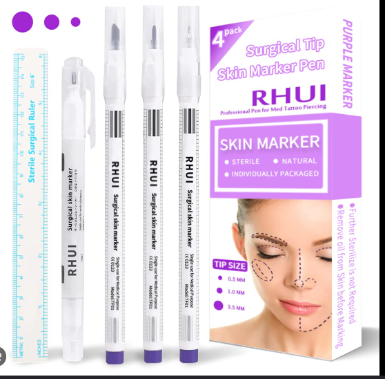
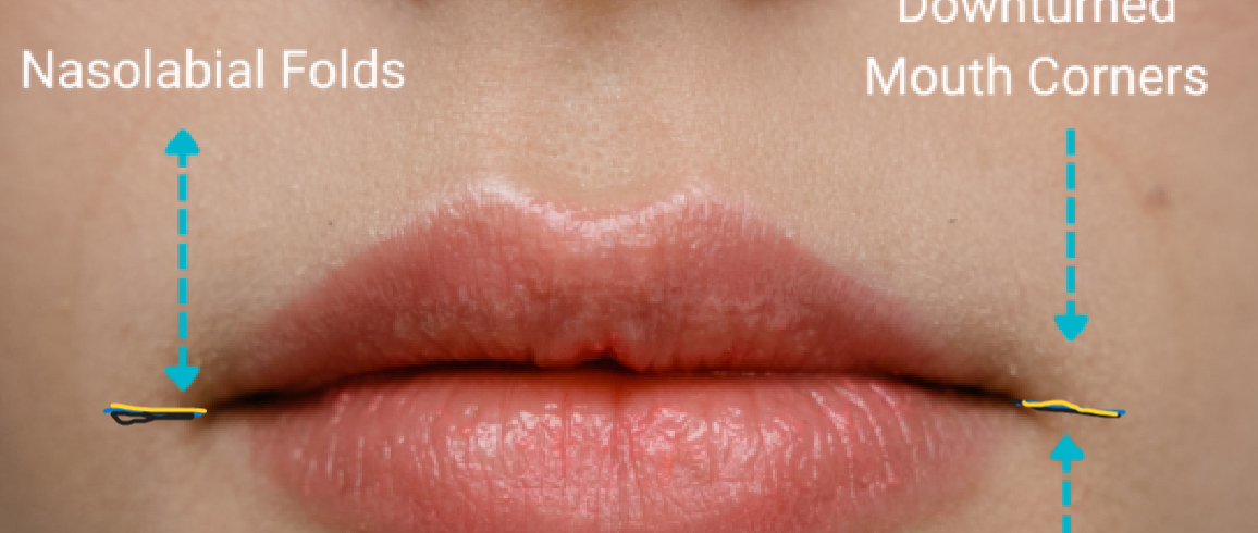
After doing ts at 1st cut only up to epi dermis and after a correct line is formed then deepen up to dermis and cut at the corner and start from where the line starts
done with ts
now use that cotton pad fresh to soak up the blood
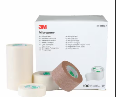
and at final use this 3m micropore surgical tape to let it heal place it vertically tho
now the question comes here is what after repeated cutting ??
At 1st mark it with surgical marker using scale mark this site only up to 1 to 2 mm length
After doing ts at 1st cut only up to epi dermis and after a correct line is formed then deepen up to dermis and cut at the corner and start from where the line starts
done with ts
now use that cotton pad fresh to soak up the blood
and at final use this 3m micropore surgical tape to let it heal place it vertically tho
now the question comes here is what after repeated cutting ??
How to manage hypertrophic fibrosis
you know what is fibrosis right ??
slop about fibrosis ( aka bulging scar line )
it just a permanent scar formation which is bright and looks not nice and is more denser than skin due to regular cutting and healing
Use tret to not get this tret goes with keratinocyte differentiation which makes the scar tissue less visible in time if you don't want scar tissue
thats all
if you want the fibrosis then just increase the cutting frequency and increase collagen
blood vessel to avoid ? = dwbi bhai you are not going deep than dermis so no need
now cut ts safely
USER WHO HAVE TRIED THE SAME PROCESS
@buccalfatremoval @Sayori @Sycophant @Navity @Hernan
you know what is fibrosis right ??
slop about fibrosis ( aka bulging scar line )
So fibrosis comes in two types localized skin fibrosis and medical fibrosis which is caused by skin condition and autoimmune diseases ,
Note : These medical fibrosis are uncontrolled and this requires a medical supervision so asking me about this won't help you .
And now clearing this stuff we can actually move on to the main topic
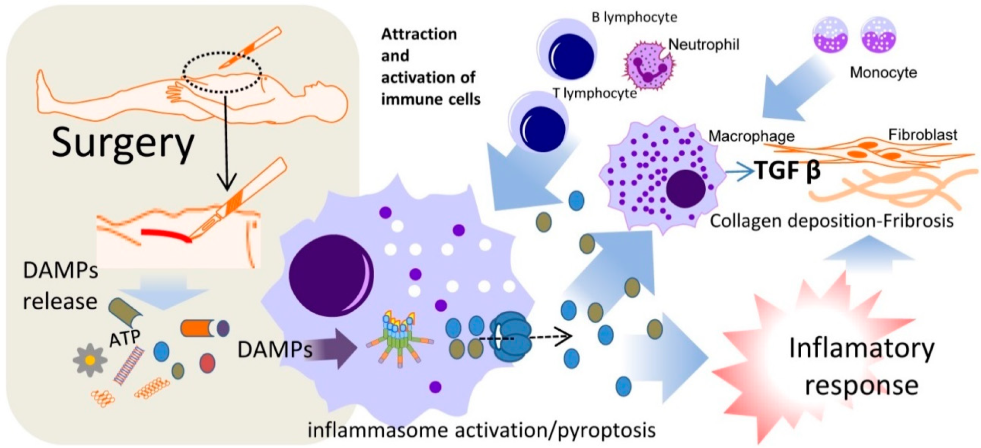
Step 1 ; at 1st the skin experiences a damage any kind of damage it may be major or minor
Damaged epithelial cells (keratinocytes) and endothelial cells (lining local blood vessels) release inflammatory molecules called damage-associated molecular patterns (DAMPs)
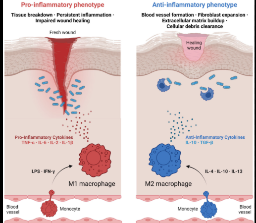
2. Influx of Profibrotic Macrophages
The cellular alarm signals recruit inflammatory white blood cells, mainly M2 macrophages.
In normal healing, M1 macrophages clean the wound and then switch to M2 cells to lay down a repair patch.
In fibrosis, these M2 macrophages stay locked in place, continuously secreting massive amounts of cytokines (cell-signaling proteins).
3. TGF-β Hyper-Activation Loop
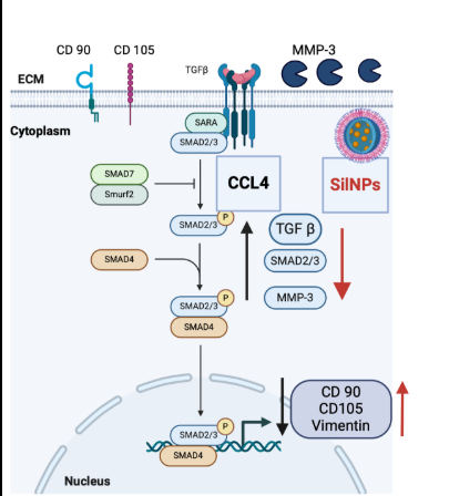
The most critical driver of fibrosis is a master-regulator protein called Transforming Growth Factor-Beta 1 (TGF-β1).
M2 macrophages and damaged cells flood the area with latent TGF-β1.
Mechanical tension in the stiffening tissue activates this latent TGF-β1.
Once active, TGF-β1 binds to receptor proteins on the surface of local resting cells, initiating an intense internal signaling cascade (primarily through the Smad2/3 pathway).
Step4 ; Phenotypic Phenotype Switch: Myofibroblast Differentiation
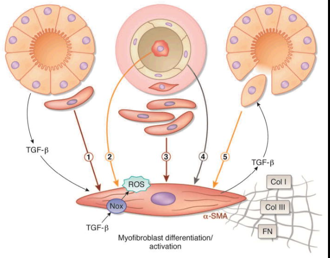
The TGF-β1 signaling forces local fibroblasts to undergo a dramatic genetic change. They differentiate into myofibroblasts.
Myofibroblasts express a protein called Alpha-Smooth Muscle Actin (α-SMA).
α-SMA gives cells internal muscle-like cables, allowing them to physically grip the surrounding tissue and pull it tightly together.
This cell contraction creates a cycle: more tissue tension → more TGF-β1 activation → more myofibroblasts.
Step5 ; Extracellular Matrix (ECM) Synthesis and Cross-Linking
The hyper-activated myofibroblasts begin synthesizing structural materials at a rate the body cannot keep up with, flooding the Extracellular Matrix (ECM).
Collagen Shift: Production of Collagen Type III drops which are replaced by massive quantitiy of rigid and dense Collagen Type I.
Cross-Linking: Cells secrete an enzyme called Lysyl Oxidase (LOX). LOX chemically cross-linking the parallel
collagen fibers tightly that the body's natural recycling enzymes MMPs can no longer break them down.
6. Structural Rigidity and Loss of Function
As the densely cross-linked Collagen Type I crowd out the normal cellular space, the microenvironment becomes choked. Blood capillaries are compressed, leading to localized hypoxia (low oxygen), which triggers even more TGF-β1 release.
Note : These medical fibrosis are uncontrolled and this requires a medical supervision so asking me about this won't help you .
And now clearing this stuff we can actually move on to the main topic
Step 1 ; at 1st the skin experiences a damage any kind of damage it may be major or minor
Damaged epithelial cells (keratinocytes) and endothelial cells (lining local blood vessels) release inflammatory molecules called damage-associated molecular patterns (DAMPs)
2. Influx of Profibrotic Macrophages
The cellular alarm signals recruit inflammatory white blood cells, mainly M2 macrophages.
In normal healing, M1 macrophages clean the wound and then switch to M2 cells to lay down a repair patch.
In fibrosis, these M2 macrophages stay locked in place, continuously secreting massive amounts of cytokines (cell-signaling proteins).
3. TGF-β Hyper-Activation Loop
The most critical driver of fibrosis is a master-regulator protein called Transforming Growth Factor-Beta 1 (TGF-β1).
M2 macrophages and damaged cells flood the area with latent TGF-β1.
Mechanical tension in the stiffening tissue activates this latent TGF-β1.
Once active, TGF-β1 binds to receptor proteins on the surface of local resting cells, initiating an intense internal signaling cascade (primarily through the Smad2/3 pathway).
Step4 ; Phenotypic Phenotype Switch: Myofibroblast Differentiation
The TGF-β1 signaling forces local fibroblasts to undergo a dramatic genetic change. They differentiate into myofibroblasts.
Myofibroblasts express a protein called Alpha-Smooth Muscle Actin (α-SMA).
α-SMA gives cells internal muscle-like cables, allowing them to physically grip the surrounding tissue and pull it tightly together.
This cell contraction creates a cycle: more tissue tension → more TGF-β1 activation → more myofibroblasts.
Step5 ; Extracellular Matrix (ECM) Synthesis and Cross-Linking
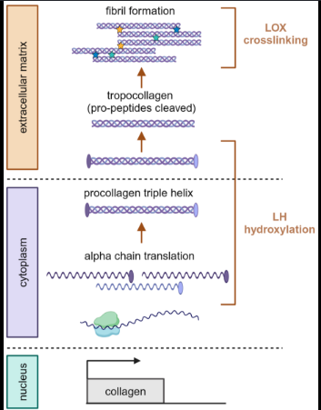
The hyper-activated myofibroblasts begin synthesizing structural materials at a rate the body cannot keep up with, flooding the Extracellular Matrix (ECM).
Collagen Shift: Production of Collagen Type III drops which are replaced by massive quantitiy of rigid and dense Collagen Type I.
Cross-Linking: Cells secrete an enzyme called Lysyl Oxidase (LOX). LOX chemically cross-linking the parallel
collagen fibers tightly that the body's natural recycling enzymes MMPs can no longer break them down.
6. Structural Rigidity and Loss of Function
As the densely cross-linked Collagen Type I crowd out the normal cellular space, the microenvironment becomes choked. Blood capillaries are compressed, leading to localized hypoxia (low oxygen), which triggers even more TGF-β1 release.
it just a permanent scar formation which is bright and looks not nice and is more denser than skin due to regular cutting and healing
Use tret to not get this tret goes with keratinocyte differentiation which makes the scar tissue less visible in time if you don't want scar tissue
thats all
if you want the fibrosis then just increase the cutting frequency and increase collagen
blood vessel to avoid ? = dwbi bhai you are not going deep than dermis so no need
now cut ts safely
USER WHO HAVE TRIED THE SAME PROCESS
@buccalfatremoval @Sayori @Sycophant @Navity @Hernan
Last edited: