





GenePilled
Iron
- Joined
- Mar 11, 2026
- Posts
- 43
- Reputation
- 23
SPIN4 (spindlin family member 4) is a gene that provides instructions for making a protein involved in regulating cellular activity and gene expression. It belongs to the spindlin family, whose proteins can interact with chromatin and help influence which genes are turned on or off. SPIN4 is not a well-established human growth gene, and there is currently limited evidence that normal variation in SPIN4 directly determines height or controls growth-plate activity.
Now Since we know SPIN4 may influence gene regulation in cells
If we inhibit spin4 we could grow faster with similar and better effects of fgfr3 inhibition with zero or less sides
Now a
A loss-of-function variant in the SPIN4 (Spindlin Family Member 4) gene causes an X-linked overgrowth syndrome characterized by extreme tall stature
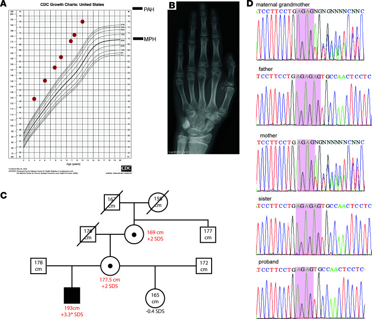
Now let’s look at a case of a boy who’s spin4 gene was gone/inactive
A) Proband’s growth chart for stature. MPH, midparental height. PAH, predicted adult height. (B) Proband’s bone age x-ray. The bone age x-ray was read as 13 years, 6 months, at chronological age 12 years, 5 months. (C) Pedigree of the family. Closed circles and rectangles represent affected subjects with height gain. Open circles and rectangles represent unaffected subjects. SDS height gain in red was calculated by subjects’ current height SDS minus subject’s midparental height SDS, except for proband’s height gain (*), which was calculated by proband’s predicted adult height SDS minus subject’s midparental height. (D) Sanger sequencing of the frameshift variant (c.312_313AGdel) in the family. MGM, maternal grandmother. AG repeats are marked in pink boxes.
Because of his excessive linear growth, he had undergone a comprehensive evaluation. His brain MRI was unremarkable except for a residual structure of Rathke’s pouch. CT of the abdomen showed increased liver and spleen size. His thyroid-stimulating hormone (TSH) and free thyroxine (T4) were normal. His IGF-1 was 179 ng/mL and testosterone was 58 ng/dL, both normal for Tanner stage 2. Repeat IGF-1 levels at different ages were also consistently unremarkable. Serum IGF-2 levels were normal in the proband and other affected family members (Supplemental Figure 1; supplemental material available online with this article; https://doi.org/10.1172/jci.insight.167074DS1). Karyotype, mutation analysis in NSD1 for Sotos syndrome, and chromosome 11p15 analysis (multiplex ligation–dependent probe amplification [MLPA] and methylation) for Beckwith-Wiedemann syndrome were normal.
On physical exam, he was noted to have protruding ears, mildly wide-spaced nipples, large flat feet, and long arms. He showed down-slanting deep-set eyes and a prominent philtrum. He had no evidence of macroglossia, joint hypermobility, scoliosis, or hemihypertrophy.
What we could get from this was he was abnormally tall for his age while not having any spinal issues unlike patients with inactive fgfr3 and nearly no symptoms
Now let’s look at figure 3
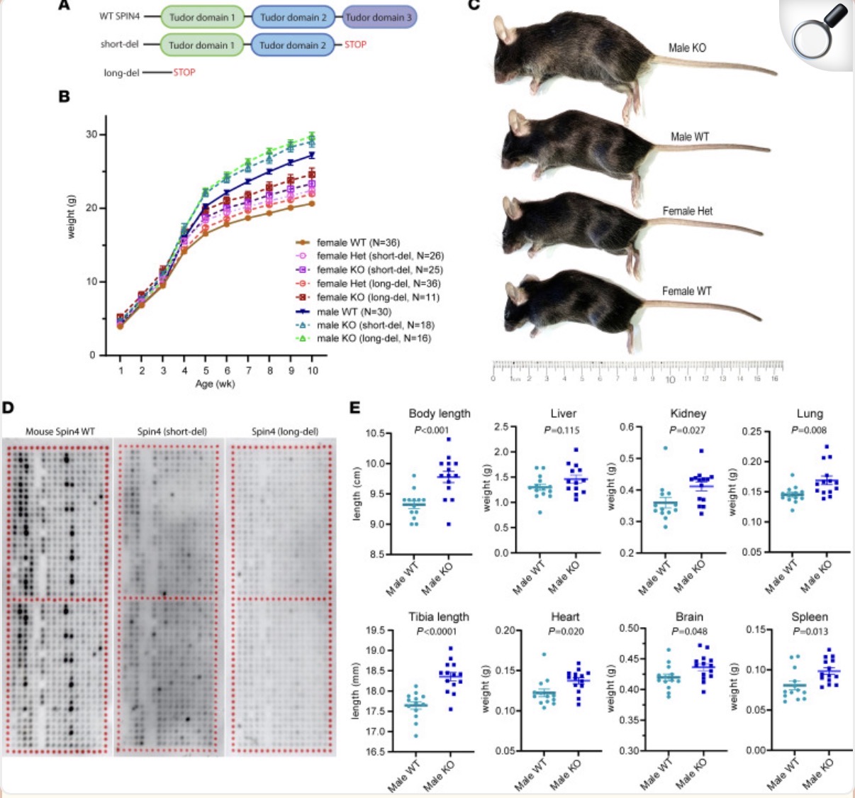
At 10 weeks of age, an increase in overall body length, tibia length, and most organ weights was observed in male KO mice compared with WT male littermates (Figure 3E). The increases in the mass of these organs was proportional to the increase in overall body mass (Supplemental Figure 2), suggesting that loss of Spin4 induces uniform overgrowth in multiple tissues. Histone arrays confirmed that both the long and short deletion of mouse Spin4 greatly diminished or abolished histone binding (Figure 3D). Our findings in the mouse models indicate that Spin4 is a negative regulator of mammalian body growth and support the hypothesis that hemizygous loss of Spin4 function contributes to the overgrowth phenotype in our patient and that heterozygous loss of Spin4 function contributes to the height gain observed in the patient’s mother and maternal grandmother.
Now somthing extremely interesting is the growth plate widening which is seen in erda use without symptoms both mice and humans with spin4 loss of function have overgrowth and are 2 deviations taller than others from these studies.
Here’s the full study because am not listing everything here but to (summarize spin4 loss of function can increases cell proliferation making you extremely taller growth plate widening which is seen in erda use and the most important part minimal side effects no scoliosis in anything and was tested in multiple mice and humans) please rep this thread as it’s my first and am new to.org this is the full study
 pmc.ncbi.nlm.nih.gov
Now Since we know SPIN4 may influence gene regulation in cells
If we inhibit spin4 we could grow faster with similar and better effects of fgfr3 inhibition with zero or less sides
Now a
A loss-of-function variant in the SPIN4 (Spindlin Family Member 4) gene causes an X-linked overgrowth syndrome characterized by extreme tall stature
Now let’s look at a case of a boy who’s spin4 gene was gone/inactive
A) Proband’s growth chart for stature. MPH, midparental height. PAH, predicted adult height. (B) Proband’s bone age x-ray. The bone age x-ray was read as 13 years, 6 months, at chronological age 12 years, 5 months. (C) Pedigree of the family. Closed circles and rectangles represent affected subjects with height gain. Open circles and rectangles represent unaffected subjects. SDS height gain in red was calculated by subjects’ current height SDS minus subject’s midparental height SDS, except for proband’s height gain (*), which was calculated by proband’s predicted adult height SDS minus subject’s midparental height. (D) Sanger sequencing of the frameshift variant (c.312_313AGdel) in the family. MGM, maternal grandmother. AG repeats are marked in pink boxes.
Because of his excessive linear growth, he had undergone a comprehensive evaluation. His brain MRI was unremarkable except for a residual structure of Rathke’s pouch. CT of the abdomen showed increased liver and spleen size. His thyroid-stimulating hormone (TSH) and free thyroxine (T4) were normal. His IGF-1 was 179 ng/mL and testosterone was 58 ng/dL, both normal for Tanner stage 2. Repeat IGF-1 levels at different ages were also consistently unremarkable. Serum IGF-2 levels were normal in the proband and other affected family members (Supplemental Figure 1; supplemental material available online with this article; https://doi.org/10.1172/jci.insight.167074DS1). Karyotype, mutation analysis in NSD1 for Sotos syndrome, and chromosome 11p15 analysis (multiplex ligation–dependent probe amplification [MLPA] and methylation) for Beckwith-Wiedemann syndrome were normal.
On physical exam, he was noted to have protruding ears, mildly wide-spaced nipples, large flat feet, and long arms. He showed down-slanting deep-set eyes and a prominent philtrum. He had no evidence of macroglossia, joint hypermobility, scoliosis, or hemihypertrophy.
What we could get from this was he was abnormally tall for his age while not having any spinal issues unlike patients with inactive fgfr3 and nearly no symptoms
Now let’s look at figure 3
Figure 3. Loss of Spin4 causes overgrowth in mouse.
A) Schematic diagram showing the location of Spin4 deletion in 2 lines of mice generated. One line with a short deletion has only Tudor domain 3 removed, while the other line with a long deletion has all 3 Tudor domains removed. (B) Growth chart showing male and female WT mice, male hemizygous (KO), and female heterozygous (Het) or homozygous (KO) in both lines. Body weight is significantly increased in both lines of mice in both males and females (2-way ANOVA for age and genotype, P < 0.01 for WT versus KO in both sexes, n = 11–36). (C) Representative image of male KO and female heterozygous mice compared with WT littermates at age 10 weeks. (D) Histone array showed that both the long and short deletion of mouse Spin4 demonstrated loss of histone peptide binding. Each spot on the array contains a peptide that represents a peptide portion of a histone that has undergone 1 or more specific posttranslational modifications. Darker color indicates greater binding of the indicated Spin protein to that modified peptide. The upper and lower halves of each array are used as technical replicates. (E) Body and tibia length and organ weights were measured in 10-week-old WT and KO (data combined from long deletion and short deletion) males. The weights of all organs except liver weight were significantly increased in the KO male. Significance was found with Student’s t test (n = 14–16).At 10 weeks of age, an increase in overall body length, tibia length, and most organ weights was observed in male KO mice compared with WT male littermates (Figure 3E). The increases in the mass of these organs was proportional to the increase in overall body mass (Supplemental Figure 2), suggesting that loss of Spin4 induces uniform overgrowth in multiple tissues. Histone arrays confirmed that both the long and short deletion of mouse Spin4 greatly diminished or abolished histone binding (Figure 3D). Our findings in the mouse models indicate that Spin4 is a negative regulator of mammalian body growth and support the hypothesis that hemizygous loss of Spin4 function contributes to the overgrowth phenotype in our patient and that heterozygous loss of Spin4 function contributes to the height gain observed in the patient’s mother and maternal grandmother.
Expanded resting zone of the growth plate in Spin4-KO mice.
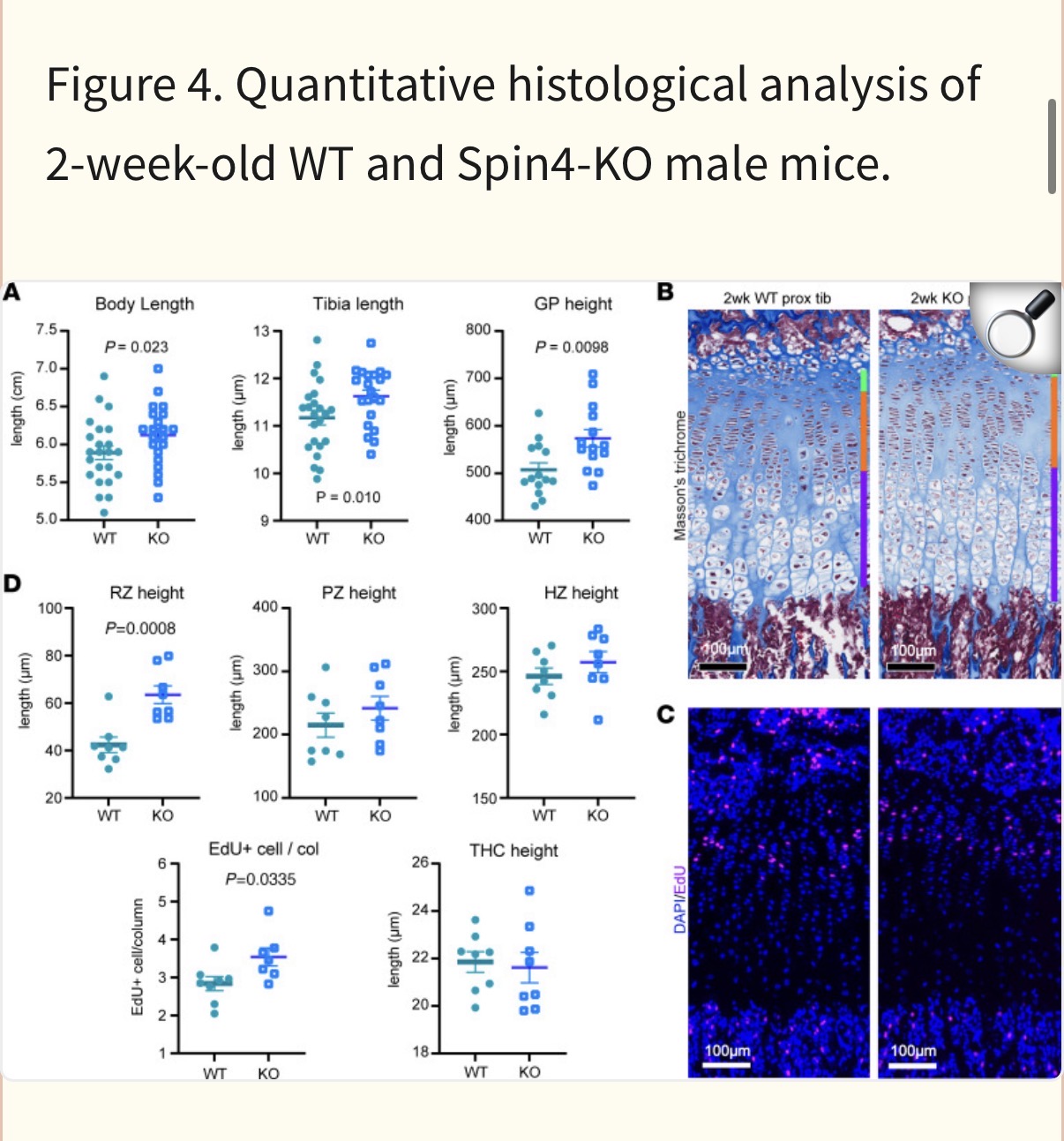
Because loss of SPIN4/Spin4 had a prominent effect on stature of the human subjects and on bone length in mice, we next focused on the growth plate, the cartilage responsible for bone elongation. We chose to study the mice at 2 weeks of age because, at this point, the secondary ossification center has formed, separating the growth plate from the articular cartilage, and skeletal growth is rapid at that age. We found that both body and tibial bone length were already significantly increased in the male KO mice compared with WT male littermates at this age (Figure 4A). In addition, 4 (liver, lung, heart, and brain) of the 6 organs studied showed increased weight (Supplemental Figure 3), suggesting that Spin4 mutation was already having a measurable impact on organ growth at 2 weeks of age (Supplemental Figure 3). Again, we assessed a possible involvement of IGF-2 in the altered body growth, but neither serum IGF-2 levels nor mRNA levels of Igf2 or H19 (measured in liver and cultured growth plate chondrocytes) showed any significant difference between male KO mice and male WT littermates at 2 weeks of ageNow somthing extremely interesting is the growth plate widening which is seen in erda use without symptoms
both mice and humans with spin4 loss of function have overgrowth and are 2 deviations taller than others from these studies.Here’s the full study because am not listing everything here but to (summarize spin4 loss of function can increases cell proliferation making you extremely taller growth plate widening
which is seen in erda use and the most important part minimal side effects no scoliosis in anything and was tested in multiple mice and humans) please rep this thread as it’s my first and am new to.org this is the full study Loss-of-function variant in SPIN4 causes an X-linked overgrowth syndrome - PMC
Overgrowth syndromes can be caused by pathogenic genetic variants in epigenetic writers, such as DNA and histone methyltransferases. However, no overgrowth disorder has previously been ascribed to variants in a gene that acts primarily as an ...
pmc.ncbi.nlm.nih.gov