





RSWT (MANLETS GTFIH)

stacylover

stacylover

Its over
Joined
Aug 13, 2025
Posts
753
Reputation
1,107
Radial shock wave therapy


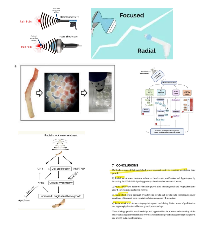
Radial shock wave therapy (RSWT) is effective because it generates extremely shortpressure pulses that propagate radially from the skin surface into the underlying tissues.These pulses are not limited to pressure alone; they produce a combination of forces,including compression, tension, shear, microstreaming in fluids, and sometimes tiny bubbles(microcavitation). This combination subjects chondrocytes and other cells to a strong andsudden mechanical shock.Cells possess specialized mechanoreceptors, such as Piezo and TRP channels, as well asintegrin clusters and focal adhesions. These receptors respond directly to changes inpressure and tension by opening calcium channels or transmitting signals to thecytoskeleton. Mechanical deformation also triggers the release of extracellular ATP, whichbinds to purinergic receptors and amplifies signaling. Within minutes, these signals convergeon central molecular pathways, including MAPK (ERK, MEK, p38), NF-κB, YAP/TAZ, RhoAGTPase, and Nrf2, whose activities can increase several-fold. This, in turn, activates geneticprograms responsible for cell proliferation, protection from apoptosis, and stimulation ofgrowth factors such as IGF-1, PTHrP, and Gli1.The ultimate result is a rapid and noticeable increase in chondrocyte proliferation andhypertrophy, thickening of the growth plate, and stimulation of longitudinal bone growth.What distinguishes RSWT is that it covers a relatively wide area and delivers a very short,complex mechanical pulse, causing cells to respond as if presented with a “strong,unavoidable signal.” This explains the potency of its effects compared to traditionalmechanical stimuli.Sources:• https://pubmed.ncbi.nlm.nih.gov/34238028/• https://stemcellres.biomedcentral.com/articles/10.1186/s13287-020-02076-w• https://pubmed.ncbi.nlm.nih.gov/37303680/

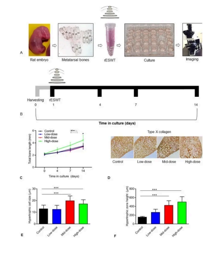
Study 1:Researchers utilized fetal rat metatarsal bones and subjected them to various frequenciesand intensities of radial extracorporeal shock wave therapy (rESWT). For our discussion,we’ll focus on the most effective parameters:• Shock Wave Parameters:• Frequency: 10 Hz• Energy: 180 mJ• Number of Pulses: 500• Session Duration: 50 secondsA single application of rESWT was administered, and the effects were assessed after 14days by measuring bone length and analyzing gene expression
Screenshot 2026 07 20 202711


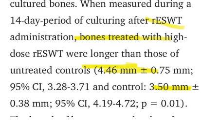
The bone length in the controlgroup was 3.50 mm ± 0.38mm, whereas in the high-dosegroup after a single applicationof rESWT, it increased to 4.46mm ± 0.75 mm, representingan approximate 27% increase.This is considered a very largeincrease, especially given thatthe treatment was applied onlyonce.Interestingly, this increase wasobserved only in the group thatreceived the high-energytreatment.
Screenshot 2026 07 20 202943


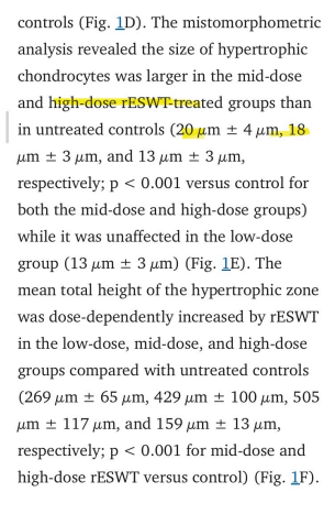
The hypertrophic chondrocyte size in thecontrol group was 13 μm ± 3 μm, whereasafter high-dose rESWT it increased to 18 μm± 3 μm, representing an approximate 38%increase.Furthermore, the mean height of thehypertrophic zone was 159 μm ± 13 μm inthe control group, and after high-doserESWT it increased to 505 μm ± 117 μm,representing an approximate 217% increase,which is considered extremely large.
Screenshot 2026 07 20 203119

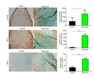
The number of proliferating cells in thegrowth plate of the control group was500 ± 412 cells, whereas afterhigh-dose rESWT it increased to 1363± 393 cells, representing anapproximate 173% increase.


Meanwhile, the number ofBcl-2–positive cells (proteins thatprotect chondrocytes from apoptosis)was 35 ± 11 cells per bone in thecontrol group, and increased to 718 ±86 cells after high-dose rESWT,representing an approximate 1950%increase, which is an extremely large increase.
1784572458732


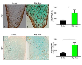
The level of NF-κB (a gene criticallyimportant for longitudinal bone growth) in thecontrol bones was 350 ± 60 positive cells perbone, whereas after high-dose rESWT itincreased to 1029 ± 262 cells, representingan approximate 194% increase.

Similarly, the level of Insulin-like GrowthFactor 1 (IGF-1), one of the most importantfactors for chondrocyte proliferation andhypertrophy, was 270 ± 121 cells in thecontrol group, and increased to 1015 ± 322cells after high-dose rESWT, representing anapproximate 276% increase.

Screenshot 2026 07 20 203630


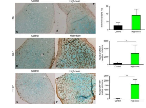
Regarding Gli1 (the glioma-associated gene 1), incontrol bones the number of positive cells was 442 ±235 cells per bone, whereas after high-dose rESWT itincreased to 2812 ± 2139 cells, representing anenormous ~536% increase. Gli1 follows Hedgehogsignaling, promoting chondrocyte proliferation andsupporting their differentiation into bone, therebydirectly contributing to longitudinal bone growth.


As for PTHrP (parathyroid hormone-related protein), inthe control group it was 21 ± 35 cells, whereas afterhigh-dose rESWT it increased to 1632 ± 493 cells, anextraordinary ~7660% increase. PTHrP functions to maintain chondrocytes in theproliferative stage, preventing their premature transition into hypertrophic cells, therebykeeping the growth plate active for a longer period and supporting increased bone length.
Screenshot 2026 07 20 203706


Interestingly, the bones exposed to high-energy rSWT were virtually protected fromchondrocyte apoptosis, largely because the proteins that safeguard chondrocytes fromprogrammed cell death, such as Bcl-2, increased by tens of times

1784572738255


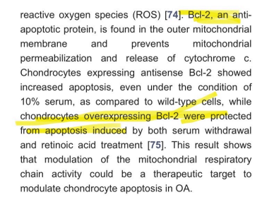
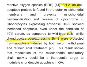
This finding is also supported byother studies, where scientistsgenetically modified chondrocytesto overexpress Bcl-2. In thesecases, the chondrocytes becameresistant to apoptosis, confirmingthe protective role of Bcl-2 againstprogrammed cell death.
Screenshot 2026 07 20 203943


Study link : https://pmc.ncbi.nlm.nih.gov/articles/PMC7145076/

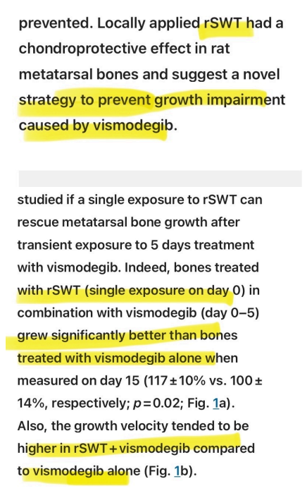
In another study on ex vivo cultured bones, the bones were exposed to a drug called vismodegib, which is a potent anti-cancer agent that strongly inhibits the Indian Hedgehog(Ihh) signaling pathway. The problem is that this pathway is critically important forlongitudinal bone growth, arguably one of the most essential factors. In children, the use of vismodegib has been associated with severe growth retardation:


> The hedgehog antagonist vismodegib is an effective anti-cancer treatment butunfortunately induces irreversible growth arrests and growth impairment, limiting its use inskeletally immature patients.

The researchers came up with an idea: why not try radial shock wave therapy (rSWT)? Theresults were truly remarkable. The study design included three groups:

1. Control group
2. Vismodegib-treated group
3. Vismodegib + single rSWT treatment group


In the vismodegib-only group, there was severe growth inhibition compared to the controlgroup. Interestingly, just one session of rSWT nearly restored growth to normal or partiallevels, effectively preventing the growth failure caused by vismodegib
1784572991856

Study link : https://www.nature.com/articles/s41598-020-69904-0


Now, let’s discuss this study, which produced truly remarkable results. The researchers used three different models, and we will focus on the first one here.


First model: cartilage discs from adolescent children

> The cartilage discs (n = 5 per patient), placed in an Eppendorf containing culture medium, were exposed to high-energy rSWT (1000 impulses, 180 mJ, 10 Hz) (Fig. 1a). After exposure to rSWT, samples were transferred back to the 24-well plate and cultured for 24 h (Fig. 1b).”

They used settings of 1000 impulses, 10 Hz, high energy 180 mJ, with a total application time of approximately 1 minute and 40 seconds. Simply put, after exposing the samples to rSWT for 1 minute and 40 seconds, they measured gene expression changes in SOX9 and COL2A1 after 24 hours.

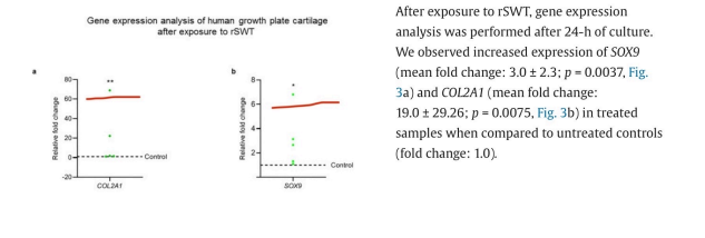
Results: The effects were literally spectacular. In one of the samples, SOX9 expressionincreased sevenfold, and COL2A1 increased nearly seventyfold. Not all samples showed such extreme results; the variability was likely due to the very short exposure time and the fact that some samples were highly sensitive to mechanical stress.

“After exposure to rSWT, gene expression analysis was performed after 24 h of culture. We observed increased expression of SOX9 (mean fold change: 3.0 ± 2.3; p = 0.0037, Fig. 3a) and COL2A1 (mean fold change: 19.0 ± 29.26; p = 0.0075, Fig. 3b) in treated samples when compared to untreated controls (fold change: 1.0).”


Interpretation: These increases are extremely meaningful. They indicate that the cartilage, instead of moving toward calcification (growth plate closure) and halting chondrocyte proliferation for ossification, was essentially reverted to a proliferative stage after just 1 minute and 40 seconds of rSWT exposure. This is truly remarkable, especially given the very short exposure time
1784573428447

Second model: 4-week-old rabbits

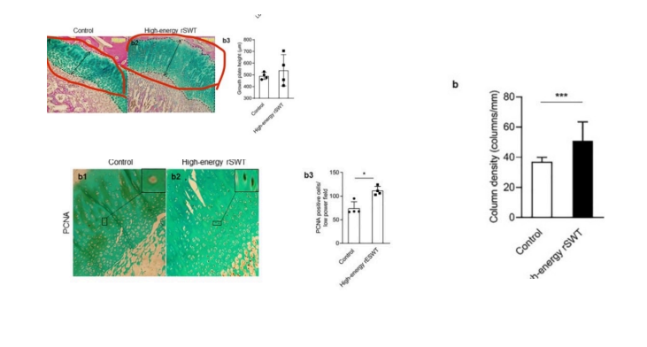
“Via a 15 mm applicator tip using ultrasound gel, animals received low-energy (1500 impulses, 90 mJ, 5 Hz, n = 4) or high-energy rSWT (3000 impulses, 180 mJ, 5 Hz, n = 4) at weekly intervals for four weeks, applied symmetrically on the medial and lateral sides of the right distal femoral growth plate, while the untreated lower limb served as a control. The more beneficial and safer dose of the two was selected for the longitudinal study. Animals were sacrificed at 14 weeks of age (4 weeks after the last rSWT dose). The distal femur was chosen as it is fast-growing and lacks an apophysis, which could have introduced errors during histomorphometric measurements. The primary outcome of the histological study was to assess cellular changes in the resting, proliferative, and hypertrophic zones after rSWT.”


Results (high-energy group only):
• The chondrocyte column density (columns per mm growth plate width) in the
proliferative zone was significantly increased in the high-energy rSWT group (51 ± 13)
compared to the control (37 ± 3; p = 0.0005).
• The height of the growth plate also increased significantly.
• Chondrocyte proliferation increased by approximately 50%.

The treatment was applied once weekly for four weeks, with each session lasting
approximately 10 minutes. Four weeks after the last treatment, results showed that
chondrocyte density in the proliferative zone increased by approximately 38%, proliferation

increased by ~50%, and the growth plate height increased noticeably.

1784573637780

Now, let’s talk about the most remarkable result of all:

Study details:

The researchers used 22-week-old rabbits and applied high-energy rSWT. The treatment
was applied to the proximal tibial growth plate. Interestingly, by 16 weeks of age, white
rabbits have already achieved about 94% of their final tibial length:

“The mean tibial length at 2 weeks was 38% of the adult length, and 94% of the adult value
was achieved by 16 weeks old “

> https://pubmed.ncbi.nlm.nih.gov/3712130/

This means that at 22 weeks, the growth plate was almost closed, with the tibia reaching
approximately 98–99% of its final length.

Remarkable finding: Despite this, just four sessions of high-energy rSWT significantly
increased tibial length, even though the treatment was applied to only one growth plate:

“At 26 weeks of age, after four sessions (at weekly intervals) of high-energy rSWT, there was
a significant increase in the differences between the lateral tibial length measured on
radiographs when compared to untreated controls (mean difference: 0.28 cm; p = 0.008).”

The average increase was 0.27 which represents approximately 7.5% growth over the
treatment period of one month:

“We found differences up to 2.7 mm after a one month treatment period. Interestingly, during the rSWT application period, up to a 7.5% growth difference was observed.”


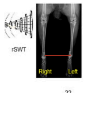
These results are truly extraordinary, especially considering that the rabbits were structurally near mature. Another important point is that mechanical stimulation applied to one limb can slightly affect the growth of the other limb, as seen in other studies ( please refer to lsjl) This means that if a second control group were measured, the effective increase could potentially reach 10%. All of these amazing effects were achieved with just four sessions, and applied to only one growth plate. Imagine the potential if rSWT were applied to multiple growth plates simultaneously! (See the figure below for a clear visual difference )

1784574206906


How to apply it (simplified explanation):

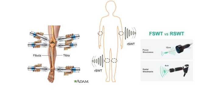
First, you need to purchase a radial shock wave therapy (rSWT) device. (See the images below to understand the difference between rSWT and FSWT.)


You can buy the device from multiple sources, preferably trusted Chinese suppliers with verified reviews. The price is approximately $150–200. (See the image below to get an idea of what it looks like.)

Settings for application:
• Frequency: 5–10 Hz
• Number of pulses: 3000
• Energy: 200-220 mJ
• Session duration: approximately 7–10 minutes-

Since we have six growth plates, we will treat one growth plate per day.

Application procedure (simplified):
1. Set the device to the parameters mentioned above.
2. Place the device over the growth plate and move it to cover the area evenly
3. Alternate between both sides to ensure the waves reach 100% of the chondrocytes.

Routine:
• Treat one growth plate per day, totaling six days per week.
• Take one day off and then repeat the routine.
• Follow this schedule, and hopefully, you will observe the remarkable results reported in the studies.


(I will include images below to show exactly how to do it.)

1784574433056


1784574448669


@Volpa
#volpamogs-
 
Last edited:
  • +1
Reactions: Absurdist, chudpiller and accinr
Radial shock wave therapy


Radial shock wave therapy (RSWT) is effective because it generates extremely shortpressure pulses that propagate radially from the skin surface into the underlying tissues.These pulses are not limited to pressure alone; they produce a combination of forces,including compression, tension, shear, microstreaming in fluids, and sometimes tiny bubbles(microcavitation). This combination subjects chondrocytes and other cells to a strong andsudden mechanical shock.Cells possess specialized mechanoreceptors, such as Piezo and TRP channels, as well asintegrin clusters and focal adhesions. These receptors respond directly to changes inpressure and tension by opening calcium channels or transmitting signals to thecytoskeleton. Mechanical deformation also triggers the release of extracellular ATP, whichbinds to purinergic receptors and amplifies signaling. Within minutes, these signals convergeon central molecular pathways, including MAPK (ERK, MEK, p38), NF-κB, YAP/TAZ, RhoAGTPase, and Nrf2, whose activities can increase several-fold. This, in turn, activates geneticprograms responsible for cell proliferation, protection from apoptosis, and stimulation ofgrowth factors such as IGF-1, PTHrP, and Gli1.The ultimate result is a rapid and noticeable increase in chondrocyte proliferation andhypertrophy, thickening of the growth plate, and stimulation of longitudinal bone growth.What distinguishes RSWT is that it covers a relatively wide area and delivers a very short,complex mechanical pulse, causing cells to respond as if presented with a “strong,unavoidable signal.” This explains the potency of its effects compared to traditionalmechanical stimuli.Sources:• https://pubmed.ncbi.nlm.nih.gov/34238028/• https://stemcellres.biomedcentral.com/articles/10.1186/s13287-020-02076-w• https://pubmed.ncbi.nlm.nih.gov/37303680/

Study 1:Researchers utilized fetal rat metatarsal bones and subjected them to various frequenciesand intensities of radial extracorporeal shock wave therapy (rESWT). For our discussion,we’ll focus on the most effective parameters:• Shock Wave Parameters:• Frequency: 10 Hz• Energy: 180 mJ• Number of Pulses: 500• Session Duration: 50 secondsA single application of rESWT was administered, and the effects were assessed after 14days by measuring bone length and analyzing gene expression


Screenshot 2026 07 20 202711


The bone length in the controlgroup was 3.50 mm ± 0.38mm, whereas in the high-dosegroup after a single applicationof rESWT, it increased to 4.46mm ± 0.75 mm, representingan approximate 27% increase.This is considered a very largeincrease, especially given thatthe treatment was applied onlyonce.Interestingly, this increase wasobserved only in the group thatreceived the high-energytreatment.

Screenshot 2026 07 20 202943


The hypertrophic chondrocyte size in thecontrol group was 13 μm ± 3 μm, whereasafter high-dose rESWT it increased to 18 μm± 3 μm, representing an approximate 38%increase.Furthermore, the mean height of thehypertrophic zone was 159 μm ± 13 μm inthe control group, and after high-doserESWT it increased to 505 μm ± 117 μm,representing an approximate 217% increase,which is considered extremely large.

Screenshot 2026 07 20 203119

The number of proliferating cells in thegrowth plate of the control group was500 ± 412 cells, whereas afterhigh-dose rESWT it increased to 1363± 393 cells, representing anapproximate 173% increase.

Meanwhile, the number ofBcl-2–positive cells (proteins thatprotect chondrocytes from apoptosis)was 35 ± 11 cells per bone in thecontrol group, and increased to 718 ±86 cells after high-dose rESWT,representing an approximate 1950%increase, which is an extremely large increase.


1784572458732

The level of NF-κB (a gene criticallyimportant for longitudinal bone growth) in thecontrol bones was 350 ± 60 positive cells perbone, whereas after high-dose rESWT itincreased to 1029 ± 262 cells, representingan approximate 194% increase.

Similarly, the level of Insulin-like GrowthFactor 1 (IGF-1), one of the most importantfactors for chondrocyte proliferation andhypertrophy, was 270 ± 121 cells in thecontrol group, and increased to 1015 ± 322cells after high-dose rESWT, representing anapproximate 276% increase.


Screenshot 2026 07 20 203630

Regarding Gli1 (the glioma-associated gene 1), incontrol bones the number of positive cells was 442 ±235 cells per bone, whereas after high-dose rESWT itincreased to 2812 ± 2139 cells, representing anenormous ~536% increase. Gli1 follows Hedgehogsignaling, promoting chondrocyte proliferation andsupporting their differentiation into bone, therebydirectly contributing to longitudinal bone growth.

As for PTHrP (parathyroid hormone-related protein), inthe control group it was 21 ± 35 cells, whereas afterhigh-dose rESWT it increased to 1632 ± 493 cells, anextraordinary ~7660% increase. PTHrP functions to maintain chondrocytes in theproliferative stage, preventing their premature transition into hypertrophic cells, therebykeeping the growth plate active for a longer period and supporting increased bone length.


Screenshot 2026 07 20 203706


Interestingly, the bones exposed to high-energy rSWT were virtually protected fromchondrocyte apoptosis, largely because the proteins that safeguard chondrocytes fromprogrammed cell death, such as Bcl-2, increased by tens of times
1784572738255


This finding is also supported byother studies, where scientistsgenetically modified chondrocytesto overexpress Bcl-2. In thesecases, the chondrocytes becameresistant to apoptosis, confirmingthe protective role of Bcl-2 againstprogrammed cell death.
1784572785128


Study link : https://pmc.ncbi.nlm.nih.gov/articles/PMC7145076/

In another study on ex vivo cultured bones, the bones were exposed to a drug called vismodegib, which is a potent anti-cancer agent that strongly inhibits the Indian Hedgehog(Ihh) signaling pathway. The problem is that this pathway is critically important forlongitudinal bone growth, arguably one of the most essential factors. In children, the use of vismodegib has been associated with severe growth retardation:

> The hedgehog antagonist vismodegib is an effective anti-cancer treatment butunfortunately induces irreversible growth arrests and growth impairment, limiting its use inskeletally immature patients.

The researchers came up with an idea: why not try radial shock wave therapy (rSWT)? Theresults were truly remarkable. The study design included three groups:

1. Control group
2. Vismodegib-treated group
3. Vismodegib + single rSWT treatment group

In the vismodegib-only group, there was severe growth inhibition compared to the controlgroup. Interestingly, just one session of rSWT nearly restored growth to normal or partiallevels, effectively preventing the growth failure caused by vismodegib


1784572991856

Study link : https://www.nature.com/articles/s41598-020-69904-0


Now, let’s discuss this study, which produced truly remarkable results. The researchers used three different models, and we will focus on the first one here.

First model: cartilage discs from adolescent children

> The cartilage discs (n = 5 per patient), placed in an Eppendorf containing culture medium, were exposed to high-energy rSWT (1000 impulses, 180 mJ, 10 Hz) (Fig. 1a). After exposure to rSWT, samples were transferred back to the 24-well plate and cultured for 24 h (Fig. 1b).”

They used settings of 1000 impulses, 10 Hz, high energy 180 mJ, with a total application time of approximately 1 minute and 40 seconds. Simply put, after exposing the samples to rSWT for 1 minute and 40 seconds, they measured gene expression changes in SOX9 and COL2A1 after 24 hours.

Results: The effects were literally spectacular. In one of the samples, SOX9 expressionincreased sevenfold, and COL2A1 increased nearly seventyfold. Not all samples showed such extreme results; the variability was likely due to the very short exposure time and the fact that some samples were highly sensitive to mechanical stress.

“After exposure to rSWT, gene expression analysis was performed after 24 h of culture. We observed increased expression of SOX9 (mean fold change: 3.0 ± 2.3; p = 0.0037, Fig. 3a) and COL2A1 (mean fold change: 19.0 ± 29.26; p = 0.0075, Fig. 3b) in treated samples when compared to untreated controls (fold change: 1.0).”

Interpretation: These increases are extremely meaningful. They indicate that the cartilage, instead of moving toward calcification (growth plate closure) and halting chondrocyte proliferation for ossification, was essentially reverted to a proliferative stage after just 1 minute and 40 seconds of rSWT exposure. This is truly remarkable, especially given the very short exposure time


1784573428447

Second model: 4-week-old rabbits

“Via a 15 mm applicator tip using ultrasound gel, animals received low-energy (1500 impulses, 90 mJ, 5 Hz, n = 4) or high-energy rSWT (3000 impulses, 180 mJ, 5 Hz, n = 4) at weekly intervals for four weeks, applied symmetrically on the medial and lateral sides of the right distal femoral growth plate, while the untreated lower limb served as a control. The more beneficial and safer dose of the two was selected for the longitudinal study. Animals were sacrificed at 14 weeks of age (4 weeks after the last rSWT dose). The distal femur was chosen as it is fast-growing and lacks an apophysis, which could have introduced errors during histomorphometric measurements. The primary outcome of the histological study was to assess cellular changes in the resting, proliferative, and hypertrophic zones after rSWT.”

Results (high-energy group only):
• The chondrocyte column density (columns per mm growth plate width) in the
proliferative zone was significantly increased in the high-energy rSWT group (51 ± 13)
compared to the control (37 ± 3; p = 0.0005).
• The height of the growth plate also increased significantly.
• Chondrocyte proliferation increased by approximately 50%.

The treatment was applied once weekly for four weeks, with each session lasting
approximately 10 minutes. Four weeks after the last treatment, results showed that
chondrocyte density in the proliferative zone increased by approximately 38%, proliferation
increased by ~50%, and the growth plate height increased noticeably.

1784573637780

Now, let’s talk about the most remarkable result of all:

Study details:
The researchers used 22-week-old rabbits and applied high-energy rSWT. The treatment
was applied to the proximal tibial growth plate. Interestingly, by 16 weeks of age, white
rabbits have already achieved about 94% of their final tibial length:

“The mean tibial length at 2 weeks was 38% of the adult length, and 94% of the adult value
was achieved by 16 weeks old “

> https://pubmed.ncbi.nlm.nih.gov/3712130/

This means that at 22 weeks, the growth plate was almost closed, with the tibia reaching
approximately 98–99% of its final length.

Remarkable finding: Despite this, just four sessions of high-energy rSWT significantly
increased tibial length, even though the treatment was applied to only one growth plate:

“At 26 weeks of age, after four sessions (at weekly intervals) of high-energy rSWT, there was
a significant increase in the differences between the lateral tibial length measured on
radiographs when compared to untreated controls (mean difference: 0.28 cm; p = 0.008).”

The average increase was 0.27 which represents approximately 7.5% growth over the
treatment period of one month:

“We found differences up to 2.7 mm after a one month treatment period. Interestingly, during the rSWT application period, up to a 7.5% growth difference was observed.”

These results are truly extraordinary, especially considering that the rabbits were structurally near mature. Another important point is that mechanical stimulation applied to one limb can slightly affect the growth of the other limb, as seen in other studies ( please refer to lsjl) This means that if a second control group were measured, the effective increase could potentially reach 10%. All of these amazing effects were achieved with just four sessions, and applied to only one growth plate. Imagine the potential if rSWT were applied to multiple growth plates simultaneously! (See the figure below for a clear visual difference )
View attachment 5391328

How to apply it (simplified explanation):

First, you need to purchase a radial shock wave therapy (rSWT) device. (See the images below to understand the difference between rSWT and FSWT.)

You can buy the device from multiple sources, preferably trusted Chinese suppliers with verified reviews. The price is approximately $150–200. (See the image below to get an idea of what it looks like.)

Settings for application:
• Frequency: 5–10 Hz
• Number of pulses: 3000
• Energy: 200-220 mJ
• Session duration: approximately 7–10 minutes-

Since we have six growth plates, we will treat one growth plate per day.

Application procedure (simplified):
1. Set the device to the parameters mentioned above.
2. Place the device over the growth plate and move it to cover the area evenly
3. Alternate between both sides to ensure the waves reach 100% of the chondrocytes.

Routine:
• Treat one growth plate per day, totaling six days per week.
• Take one day off and then repeat the routine.
• Follow this schedule, and hopefully, you will observe the remarkable results reported in the studies.

(I will include images below to show exactly how to do it.)
View attachment 5391346

View attachment 5391348
bumped for effort
mirin content
brutal that chad just wakes up and never has to come across any of this
 
  • +1
Reactions: stacylover and Absurdist
Radial shock wave therapy


Radial shock wave therapy (RSWT) is effective because it generates extremely shortpressure pulses that propagate radially from the skin surface into the underlying tissues.These pulses are not limited to pressure alone; they produce a combination of forces,including compression, tension, shear, microstreaming in fluids, and sometimes tiny bubbles(microcavitation). This combination subjects chondrocytes and other cells to a strong andsudden mechanical shock.Cells possess specialized mechanoreceptors, such as Piezo and TRP channels, as well asintegrin clusters and focal adhesions. These receptors respond directly to changes inpressure and tension by opening calcium channels or transmitting signals to thecytoskeleton. Mechanical deformation also triggers the release of extracellular ATP, whichbinds to purinergic receptors and amplifies signaling. Within minutes, these signals convergeon central molecular pathways, including MAPK (ERK, MEK, p38), NF-κB, YAP/TAZ, RhoAGTPase, and Nrf2, whose activities can increase several-fold. This, in turn, activates geneticprograms responsible for cell proliferation, protection from apoptosis, and stimulation ofgrowth factors such as IGF-1, PTHrP, and Gli1.The ultimate result is a rapid and noticeable increase in chondrocyte proliferation andhypertrophy, thickening of the growth plate, and stimulation of longitudinal bone growth.What distinguishes RSWT is that it covers a relatively wide area and delivers a very short,complex mechanical pulse, causing cells to respond as if presented with a “strong,unavoidable signal.” This explains the potency of its effects compared to traditionalmechanical stimuli.Sources:• https://pubmed.ncbi.nlm.nih.gov/34238028/• https://stemcellres.biomedcentral.com/articles/10.1186/s13287-020-02076-w• https://pubmed.ncbi.nlm.nih.gov/37303680/

Study 1:Researchers utilized fetal rat metatarsal bones and subjected them to various frequenciesand intensities of radial extracorporeal shock wave therapy (rESWT). For our discussion,we’ll focus on the most effective parameters:• Shock Wave Parameters:• Frequency: 10 Hz• Energy: 180 mJ• Number of Pulses: 500• Session Duration: 50 secondsA single application of rESWT was administered, and the effects were assessed after 14days by measuring bone length and analyzing gene expression


Screenshot 2026 07 20 202711


The bone length in the controlgroup was 3.50 mm ± 0.38mm, whereas in the high-dosegroup after a single applicationof rESWT, it increased to 4.46mm ± 0.75 mm, representingan approximate 27% increase.This is considered a very largeincrease, especially given thatthe treatment was applied onlyonce.Interestingly, this increase wasobserved only in the group thatreceived the high-energytreatment.

Screenshot 2026 07 20 202943


The hypertrophic chondrocyte size in thecontrol group was 13 μm ± 3 μm, whereasafter high-dose rESWT it increased to 18 μm± 3 μm, representing an approximate 38%increase.Furthermore, the mean height of thehypertrophic zone was 159 μm ± 13 μm inthe control group, and after high-doserESWT it increased to 505 μm ± 117 μm,representing an approximate 217% increase,which is considered extremely large.

Screenshot 2026 07 20 203119

The number of proliferating cells in thegrowth plate of the control group was500 ± 412 cells, whereas afterhigh-dose rESWT it increased to 1363± 393 cells, representing anapproximate 173% increase.

Meanwhile, the number ofBcl-2–positive cells (proteins thatprotect chondrocytes from apoptosis)was 35 ± 11 cells per bone in thecontrol group, and increased to 718 ±86 cells after high-dose rESWT,representing an approximate 1950%increase, which is an extremely large increase.

1784572458732


The level of NF-κB (a gene criticallyimportant for longitudinal bone growth) in thecontrol bones was 350 ± 60 positive cells perbone, whereas after high-dose rESWT itincreased to 1029 ± 262 cells, representingan approximate 194% increase.

Similarly, the level of Insulin-like GrowthFactor 1 (IGF-1), one of the most importantfactors for chondrocyte proliferation andhypertrophy, was 270 ± 121 cells in thecontrol group, and increased to 1015 ± 322cells after high-dose rESWT, representing anapproximate 276% increase.

Screenshot 2026 07 20 203630


Regarding Gli1 (the glioma-associated gene 1), incontrol bones the number of positive cells was 442 ±235 cells per bone, whereas after high-dose rESWT itincreased to 2812 ± 2139 cells, representing anenormous ~536% increase. Gli1 follows Hedgehogsignaling, promoting chondrocyte proliferation andsupporting their differentiation into bone, therebydirectly contributing to longitudinal bone growth.

As for PTHrP (parathyroid hormone-related protein), inthe control group it was 21 ± 35 cells, whereas afterhigh-dose rESWT it increased to 1632 ± 493 cells, anextraordinary ~7660% increase. PTHrP functions to maintain chondrocytes in theproliferative stage, preventing their premature transition into hypertrophic cells, therebykeeping the growth plate active for a longer period and supporting increased bone length.


Screenshot 2026 07 20 203706


Interestingly, the bones exposed to high-energy rSWT were virtually protected fromchondrocyte apoptosis, largely because the proteins that safeguard chondrocytes fromprogrammed cell death, such as Bcl-2, increased by tens of times
1784572738255


This finding is also supported byother studies, where scientistsgenetically modified chondrocytesto overexpress Bcl-2. In thesecases, the chondrocytes becameresistant to apoptosis, confirmingthe protective role of Bcl-2 againstprogrammed cell death.

View attachment 5391372


Study link : https://pmc.ncbi.nlm.nih.gov/articles/PMC7145076/

In another study on ex vivo cultured bones, the bones were exposed to a drug called vismodegib, which is a potent anti-cancer agent that strongly inhibits the Indian Hedgehog(Ihh) signaling pathway. The problem is that this pathway is critically important forlongitudinal bone growth, arguably one of the most essential factors. In children, the use of vismodegib has been associated with severe growth retardation:

> The hedgehog antagonist vismodegib is an effective anti-cancer treatment butunfortunately induces irreversible growth arrests and growth impairment, limiting its use inskeletally immature patients.

The researchers came up with an idea: why not try radial shock wave therapy (rSWT)? Theresults were truly remarkable. The study design included three groups:

1. Control group
2. Vismodegib-treated group
3. Vismodegib + single rSWT treatment group

In the vismodegib-only group, there was severe growth inhibition compared to the controlgroup. Interestingly, just one session of rSWT nearly restored growth to normal or partiallevels, effectively preventing the growth failure caused by vismodegib


1784572991856

Study link : https://www.nature.com/articles/s41598-020-69904-0


Now, let’s discuss this study, which produced truly remarkable results. The researchers used three different models, and we will focus on the first one here.

First model: cartilage discs from adolescent children

> The cartilage discs (n = 5 per patient), placed in an Eppendorf containing culture medium, were exposed to high-energy rSWT (1000 impulses, 180 mJ, 10 Hz) (Fig. 1a). After exposure to rSWT, samples were transferred back to the 24-well plate and cultured for 24 h (Fig. 1b).”

They used settings of 1000 impulses, 10 Hz, high energy 180 mJ, with a total application time of approximately 1 minute and 40 seconds. Simply put, after exposing the samples to rSWT for 1 minute and 40 seconds, they measured gene expression changes in SOX9 and COL2A1 after 24 hours.

Results: The effects were literally spectacular. In one of the samples, SOX9 expressionincreased sevenfold, and COL2A1 increased nearly seventyfold. Not all samples showed such extreme results; the variability was likely due to the very short exposure time and the fact that some samples were highly sensitive to mechanical stress.

“After exposure to rSWT, gene expression analysis was performed after 24 h of culture. We observed increased expression of SOX9 (mean fold change: 3.0 ± 2.3; p = 0.0037, Fig. 3a) and COL2A1 (mean fold change: 19.0 ± 29.26; p = 0.0075, Fig. 3b) in treated samples when compared to untreated controls (fold change: 1.0).”

Interpretation: These increases are extremely meaningful. They indicate that the cartilage, instead of moving toward calcification (growth plate closure) and halting chondrocyte proliferation for ossification, was essentially reverted to a proliferative stage after just 1 minute and 40 seconds of rSWT exposure. This is truly remarkable, especially given the very short exposure time


1784573428447

Second model: 4-week-old rabbits

“Via a 15 mm applicator tip using ultrasound gel, animals received low-energy (1500 impulses, 90 mJ, 5 Hz, n = 4) or high-energy rSWT (3000 impulses, 180 mJ, 5 Hz, n = 4) at weekly intervals for four weeks, applied symmetrically on the medial and lateral sides of the right distal femoral growth plate, while the untreated lower limb served as a control. The more beneficial and safer dose of the two was selected for the longitudinal study. Animals were sacrificed at 14 weeks of age (4 weeks after the last rSWT dose). The distal femur was chosen as it is fast-growing and lacks an apophysis, which could have introduced errors during histomorphometric measurements. The primary outcome of the histological study was to assess cellular changes in the resting, proliferative, and hypertrophic zones after rSWT.”

Results (high-energy group only):
• The chondrocyte column density (columns per mm growth plate width) in the
proliferative zone was significantly increased in the high-energy rSWT group (51 ± 13)
compared to the control (37 ± 3; p = 0.0005).
• The height of the growth plate also increased significantly.
• Chondrocyte proliferation increased by approximately 50%.

The treatment was applied once weekly for four weeks, with each session lasting
approximately 10 minutes. Four weeks after the last treatment, results showed that
chondrocyte density in the proliferative zone increased by approximately 38%, proliferation
increased by ~50%, and the growth plate height increased noticeably.


1784573637780

Now, let’s talk about the most remarkable result of all:

Study details:
The researchers used 22-week-old rabbits and applied high-energy rSWT. The treatment
was applied to the proximal tibial growth plate. Interestingly, by 16 weeks of age, white
rabbits have already achieved about 94% of their final tibial length:

“The mean tibial length at 2 weeks was 38% of the adult length, and 94% of the adult value
was achieved by 16 weeks old “

> https://pubmed.ncbi.nlm.nih.gov/3712130/

This means that at 22 weeks, the growth plate was almost closed, with the tibia reaching
approximately 98–99% of its final length.

Remarkable finding: Despite this, just four sessions of high-energy rSWT significantly
increased tibial length, even though the treatment was applied to only one growth plate:

“At 26 weeks of age, after four sessions (at weekly intervals) of high-energy rSWT, there was
a significant increase in the differences between the lateral tibial length measured on
radiographs when compared to untreated controls (mean difference: 0.28 cm; p = 0.008).”

The average increase was 0.27 which represents approximately 7.5% growth over the
treatment period of one month:

“We found differences up to 2.7 mm after a one month treatment period. Interestingly, during the rSWT application period, up to a 7.5% growth difference was observed.”

These results are truly extraordinary, especially considering that the rabbits were structurally near mature. Another important point is that mechanical stimulation applied to one limb can slightly affect the growth of the other limb, as seen in other studies ( please refer to lsjl) This means that if a second control group were measured, the effective increase could potentially reach 10%. All of these amazing effects were achieved with just four sessions, and applied to only one growth plate. Imagine the potential if rSWT were applied to multiple growth plates simultaneously! (See the figure below for a clear visual difference )

View attachment 5391328

How to apply it (simplified explanation):

First, you need to purchase a radial shock wave therapy (rSWT) device. (See the images below to understand the difference between rSWT and FSWT.)

You can buy the device from multiple sources, preferably trusted Chinese suppliers with verified reviews. The price is approximately $150–200. (See the image below to get an idea of what it looks like.)

Settings for application:
• Frequency: 5–10 Hz
• Number of pulses: 3000
• Energy: 200-220 mJ
• Session duration: approximately 7–10 minutes-

Since we have six growth plates, we will treat one growth plate per day.

Application procedure (simplified):
1. Set the device to the parameters mentioned above.
2. Place the device over the growth plate and move it to cover the area evenly
3. Alternate between both sides to ensure the waves reach 100% of the chondrocytes.

Routine:
• Treat one growth plate per day, totaling six days per week.
• Take one day off and then repeat the routine.
• Follow this schedule, and hopefully, you will observe the remarkable results reported in the studies.

(I will include images below to show exactly how to do it.)
View attachment 5391346

View attachment 5391348


@Volpa
#volpamogs-
dnr now but will read later cause im a manlet.
 
  • +1
Reactions: stacylover
Radial shock wave therapy


Radial shock wave therapy (RSWT) is effective because it generates extremely shortpressure pulses that propagate radially from the skin surface into the underlying tissues.These pulses are not limited to pressure alone; they produce a combination of forces,including compression, tension, shear, microstreaming in fluids, and sometimes tiny bubbles(microcavitation). This combination subjects chondrocytes and other cells to a strong andsudden mechanical shock.Cells possess specialized mechanoreceptors, such as Piezo and TRP channels, as well asintegrin clusters and focal adhesions. These receptors respond directly to changes inpressure and tension by opening calcium channels or transmitting signals to thecytoskeleton. Mechanical deformation also triggers the release of extracellular ATP, whichbinds to purinergic receptors and amplifies signaling. Within minutes, these signals convergeon central molecular pathways, including MAPK (ERK, MEK, p38), NF-κB, YAP/TAZ, RhoAGTPase, and Nrf2, whose activities can increase several-fold. This, in turn, activates geneticprograms responsible for cell proliferation, protection from apoptosis, and stimulation ofgrowth factors such as IGF-1, PTHrP, and Gli1.The ultimate result is a rapid and noticeable increase in chondrocyte proliferation andhypertrophy, thickening of the growth plate, and stimulation of longitudinal bone growth.What distinguishes RSWT is that it covers a relatively wide area and delivers a very short,complex mechanical pulse, causing cells to respond as if presented with a “strong,unavoidable signal.” This explains the potency of its effects compared to traditionalmechanical stimuli.Sources:• https://pubmed.ncbi.nlm.nih.gov/34238028/• https://stemcellres.biomedcentral.com/articles/10.1186/s13287-020-02076-w• https://pubmed.ncbi.nlm.nih.gov/37303680/

Study 1:Researchers utilized fetal rat metatarsal bones and subjected them to various frequenciesand intensities of radial extracorporeal shock wave therapy (rESWT). For our discussion,we’ll focus on the most effective parameters:• Shock Wave Parameters:• Frequency: 10 Hz• Energy: 180 mJ• Number of Pulses: 500• Session Duration: 50 secondsA single application of rESWT was administered, and the effects were assessed after 14days by measuring bone length and analyzing gene expression


Screenshot 2026 07 20 202711


The bone length in the controlgroup was 3.50 mm ± 0.38mm, whereas in the high-dosegroup after a single applicationof rESWT, it increased to 4.46mm ± 0.75 mm, representingan approximate 27% increase.This is considered a very largeincrease, especially given thatthe treatment was applied onlyonce.Interestingly, this increase wasobserved only in the group thatreceived the high-energytreatment.

Screenshot 2026 07 20 202943


The hypertrophic chondrocyte size in thecontrol group was 13 μm ± 3 μm, whereasafter high-dose rESWT it increased to 18 μm± 3 μm, representing an approximate 38%increase.Furthermore, the mean height of thehypertrophic zone was 159 μm ± 13 μm inthe control group, and after high-doserESWT it increased to 505 μm ± 117 μm,representing an approximate 217% increase,which is considered extremely large.

Screenshot 2026 07 20 203119

The number of proliferating cells in thegrowth plate of the control group was500 ± 412 cells, whereas afterhigh-dose rESWT it increased to 1363± 393 cells, representing anapproximate 173% increase.

Meanwhile, the number ofBcl-2–positive cells (proteins thatprotect chondrocytes from apoptosis)was 35 ± 11 cells per bone in thecontrol group, and increased to 718 ±86 cells after high-dose rESWT,representing an approximate 1950%increase, which is an extremely large increase.

1784572458732


The level of NF-κB (a gene criticallyimportant for longitudinal bone growth) in thecontrol bones was 350 ± 60 positive cells perbone, whereas after high-dose rESWT itincreased to 1029 ± 262 cells, representingan approximate 194% increase.

Similarly, the level of Insulin-like GrowthFactor 1 (IGF-1), one of the most importantfactors for chondrocyte proliferation andhypertrophy, was 270 ± 121 cells in thecontrol group, and increased to 1015 ± 322cells after high-dose rESWT, representing anapproximate 276% increase.

Screenshot 2026 07 20 203630


Regarding Gli1 (the glioma-associated gene 1), incontrol bones the number of positive cells was 442 ±235 cells per bone, whereas after high-dose rESWT itincreased to 2812 ± 2139 cells, representing anenormous ~536% increase. Gli1 follows Hedgehogsignaling, promoting chondrocyte proliferation andsupporting their differentiation into bone, therebydirectly contributing to longitudinal bone growth.

As for PTHrP (parathyroid hormone-related protein), inthe control group it was 21 ± 35 cells, whereas afterhigh-dose rESWT it increased to 1632 ± 493 cells, anextraordinary ~7660% increase. PTHrP functions to maintain chondrocytes in theproliferative stage, preventing their premature transition into hypertrophic cells, therebykeeping the growth plate active for a longer period and supporting increased bone length.


Screenshot 2026 07 20 203706


Interestingly, the bones exposed to high-energy rSWT were virtually protected fromchondrocyte apoptosis, largely because the proteins that safeguard chondrocytes fromprogrammed cell death, such as Bcl-2, increased by tens of times
1784572738255


This finding is also supported byother studies, where scientistsgenetically modified chondrocytesto overexpress Bcl-2. In thesecases, the chondrocytes becameresistant to apoptosis, confirmingthe protective role of Bcl-2 againstprogrammed cell death.

View attachment 5391372


Study link : https://pmc.ncbi.nlm.nih.gov/articles/PMC7145076/

In another study on ex vivo cultured bones, the bones were exposed to a drug called vismodegib, which is a potent anti-cancer agent that strongly inhibits the Indian Hedgehog(Ihh) signaling pathway. The problem is that this pathway is critically important forlongitudinal bone growth, arguably one of the most essential factors. In children, the use of vismodegib has been associated with severe growth retardation:

> The hedgehog antagonist vismodegib is an effective anti-cancer treatment butunfortunately induces irreversible growth arrests and growth impairment, limiting its use inskeletally immature patients.

The researchers came up with an idea: why not try radial shock wave therapy (rSWT)? Theresults were truly remarkable. The study design included three groups:

1. Control group
2. Vismodegib-treated group
3. Vismodegib + single rSWT treatment group

In the vismodegib-only group, there was severe growth inhibition compared to the controlgroup. Interestingly, just one session of rSWT nearly restored growth to normal or partiallevels, effectively preventing the growth failure caused by vismodegib


1784572991856

Study link : https://www.nature.com/articles/s41598-020-69904-0


Now, let’s discuss this study, which produced truly remarkable results. The researchers used three different models, and we will focus on the first one here.

First model: cartilage discs from adolescent children

> The cartilage discs (n = 5 per patient), placed in an Eppendorf containing culture medium, were exposed to high-energy rSWT (1000 impulses, 180 mJ, 10 Hz) (Fig. 1a). After exposure to rSWT, samples were transferred back to the 24-well plate and cultured for 24 h (Fig. 1b).”

They used settings of 1000 impulses, 10 Hz, high energy 180 mJ, with a total application time of approximately 1 minute and 40 seconds. Simply put, after exposing the samples to rSWT for 1 minute and 40 seconds, they measured gene expression changes in SOX9 and COL2A1 after 24 hours.

Results: The effects were literally spectacular. In one of the samples, SOX9 expressionincreased sevenfold, and COL2A1 increased nearly seventyfold. Not all samples showed such extreme results; the variability was likely due to the very short exposure time and the fact that some samples were highly sensitive to mechanical stress.

“After exposure to rSWT, gene expression analysis was performed after 24 h of culture. We observed increased expression of SOX9 (mean fold change: 3.0 ± 2.3; p = 0.0037, Fig. 3a) and COL2A1 (mean fold change: 19.0 ± 29.26; p = 0.0075, Fig. 3b) in treated samples when compared to untreated controls (fold change: 1.0).”

Interpretation: These increases are extremely meaningful. They indicate that the cartilage, instead of moving toward calcification (growth plate closure) and halting chondrocyte proliferation for ossification, was essentially reverted to a proliferative stage after just 1 minute and 40 seconds of rSWT exposure. This is truly remarkable, especially given the very short exposure time


1784573428447

Second model: 4-week-old rabbits

“Via a 15 mm applicator tip using ultrasound gel, animals received low-energy (1500 impulses, 90 mJ, 5 Hz, n = 4) or high-energy rSWT (3000 impulses, 180 mJ, 5 Hz, n = 4) at weekly intervals for four weeks, applied symmetrically on the medial and lateral sides of the right distal femoral growth plate, while the untreated lower limb served as a control. The more beneficial and safer dose of the two was selected for the longitudinal study. Animals were sacrificed at 14 weeks of age (4 weeks after the last rSWT dose). The distal femur was chosen as it is fast-growing and lacks an apophysis, which could have introduced errors during histomorphometric measurements. The primary outcome of the histological study was to assess cellular changes in the resting, proliferative, and hypertrophic zones after rSWT.”

Results (high-energy group only):
• The chondrocyte column density (columns per mm growth plate width) in the
proliferative zone was significantly increased in the high-energy rSWT group (51 ± 13)
compared to the control (37 ± 3; p = 0.0005).
• The height of the growth plate also increased significantly.
• Chondrocyte proliferation increased by approximately 50%.

The treatment was applied once weekly for four weeks, with each session lasting
approximately 10 minutes. Four weeks after the last treatment, results showed that
chondrocyte density in the proliferative zone increased by approximately 38%, proliferation
increased by ~50%, and the growth plate height increased noticeably.


1784573637780

Now, let’s talk about the most remarkable result of all:

Study details:
The researchers used 22-week-old rabbits and applied high-energy rSWT. The treatment
was applied to the proximal tibial growth plate. Interestingly, by 16 weeks of age, white
rabbits have already achieved about 94% of their final tibial length:

“The mean tibial length at 2 weeks was 38% of the adult length, and 94% of the adult value
was achieved by 16 weeks old “

> https://pubmed.ncbi.nlm.nih.gov/3712130/

This means that at 22 weeks, the growth plate was almost closed, with the tibia reaching
approximately 98–99% of its final length.

Remarkable finding: Despite this, just four sessions of high-energy rSWT significantly
increased tibial length, even though the treatment was applied to only one growth plate:

“At 26 weeks of age, after four sessions (at weekly intervals) of high-energy rSWT, there was
a significant increase in the differences between the lateral tibial length measured on
radiographs when compared to untreated controls (mean difference: 0.28 cm; p = 0.008).”

The average increase was 0.27 which represents approximately 7.5% growth over the
treatment period of one month:

“We found differences up to 2.7 mm after a one month treatment period. Interestingly, during the rSWT application period, up to a 7.5% growth difference was observed.”

These results are truly extraordinary, especially considering that the rabbits were structurally near mature. Another important point is that mechanical stimulation applied to one limb can slightly affect the growth of the other limb, as seen in other studies ( please refer to lsjl) This means that if a second control group were measured, the effective increase could potentially reach 10%. All of these amazing effects were achieved with just four sessions, and applied to only one growth plate. Imagine the potential if rSWT were applied to multiple growth plates simultaneously! (See the figure below for a clear visual difference )

View attachment 5391328

How to apply it (simplified explanation):

First, you need to purchase a radial shock wave therapy (rSWT) device. (See the images below to understand the difference between rSWT and FSWT.)

You can buy the device from multiple sources, preferably trusted Chinese suppliers with verified reviews. The price is approximately $150–200. (See the image below to get an idea of what it looks like.)

Settings for application:
• Frequency: 5–10 Hz
• Number of pulses: 3000
• Energy: 200-220 mJ
• Session duration: approximately 7–10 minutes-

Since we have six growth plates, we will treat one growth plate per day.

Application procedure (simplified):
1. Set the device to the parameters mentioned above.
2. Place the device over the growth plate and move it to cover the area evenly
3. Alternate between both sides to ensure the waves reach 100% of the chondrocytes.

Routine:
• Treat one growth plate per day, totaling six days per week.
• Take one day off and then repeat the routine.
• Follow this schedule, and hopefully, you will observe the remarkable results reported in the studies.

(I will include images below to show exactly how to do it.)

View attachment 5391346

View attachment 5391348


@Volpa
#volpamogs-
which nootropic did you use to right this? also you should really make the formatting easier to read its hard to look at.
 
  • +1
Reactions: stacylover
which nootropic did you use to right this? also you should really make the formatting easier to read its hard to look at.
I can edit it dm me how I want to reach botb
 

Similar threads

stacylover
Replies
1
Views
19
stacylover
stacylover
stacylover
Replies
2
Views
18
KellSS
KellSS
stacylover
Replies
0
Views
41
stacylover
stacylover
stacylover
Replies
9
Views
95
stacylover
stacylover
uglyretardednigga
Replies
11
Views
282
larpsahurcel
larpsahurcel

Users who are viewing this thread

  • knut1
Back
Top
Sponsored
Stake.us
America's #1 Social Casino
Slots, Poker & More
Join Now →