




SaintSlep
Innocent little boy tainted by female nature
- Joined
- Feb 17, 2026
- Posts
- 161
- Reputation
- 120
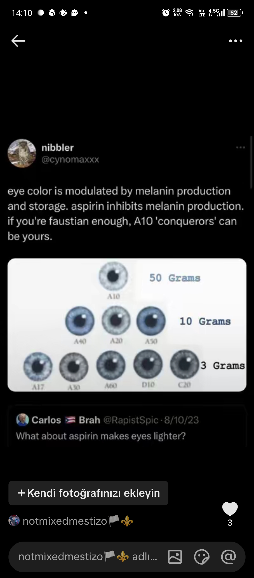
THE COMPLETE THEORY FOR ASPIRIN CHANGING IRIS COLOR. (reversing iris pigment to genetic baseline)
The Hypothesis : Systemic administration of acetylsalicylic acid (aspirin) down-regulates iris hyperpigmentation by inhibiting cyclooxygenase (COX) pathways, thereby decreasing the downstream inflammatory signaling that stimulates melanogenesis in uveal melanocytes.
Where this fits: When the iris experiences trauma or heavy UV exposure, it triggers a localized inflammatory response. This response causes a massive spike in prostaglandins and Alpha-MSH right inside the eye. The "Alarm" State: These chemicals act as an alarm system, forcing iris melanocytes out of their usual "dormant" state to start actively producing extra melanin as a protective shield. This is how you get post-inflammatory hyperpigmentation or dark spots on the iris.
Where Aspirin Fits: By introducing aspirin, you are systemically shutting down the COX enzymes producing those prostaglandins and blocking the Alpha-MSH signal. You are essentially turning off the "alarm." Without the alarm, the cellular signal to produce excess melanin drops to zero.
Pathways in which aspirin works: Alpha msh: inhibits α-MSH ↓
Localized injury/trauma response ↓
alters blood-aqueous barrier ↓
spikes α-MSH and forces COX enzymes to produce prostaglandins.aspirin inhibits COX enzymes ↓
decreases production of prostaglandins ↓
downregulates master transcription factor MITF ↓
suppresses rate-limiting enzyme tyrosinase ↓
halts synthesis of eumelanin pigment ↓
causes retraction and atrophy of melanocyte dendrites ↓
blocks physical transfer of melanosomes to iris stromal cells ↓
reduces excess melanin within the iris ↓
allows ocular macrophages (immune cleanup cells) to clear away existing excess pigment without new pigment replacing it.
Sources/Literature: (Used AI overview )
)
The Hypothesis
: Systemic administration of acetylsalicylic acid (aspirin) down-regulates iris hyperpigmentation by inhibiting cyclooxygenase (COX) pathways, thereby decreasing the downstream inflammatory signaling that stimulates melanogenesis in uveal melanocytes.Where this fits
: When the iris experiences trauma or heavy UV exposure, it triggers a localized inflammatory response. This response causes a massive spike in prostaglandins and Alpha-MSH right inside the eye. The "Alarm" State: These chemicals act as an alarm system, forcing iris melanocytes out of their usual "dormant" state to start actively producing extra melanin as a protective shield. This is how you get post-inflammatory hyperpigmentation or dark spots on the iris.Where Aspirin Fits
: By introducing aspirin, you are systemically shutting down the COX enzymes producing those prostaglandins and blocking the Alpha-MSH signal. You are essentially turning off the "alarm." Without the alarm, the cellular signal to produce excess melanin drops to zero.Pathways in which aspirin works
: Alpha msh: inhibits α-MSH ↓Localized injury/trauma response ↓
alters blood-aqueous barrier ↓
spikes α-MSH and forces COX enzymes to produce prostaglandins.aspirin inhibits COX enzymes ↓
decreases production of prostaglandins ↓
downregulates master transcription factor MITF ↓
suppresses rate-limiting enzyme tyrosinase ↓
halts synthesis of eumelanin pigment ↓
causes retraction and atrophy of melanocyte dendrites ↓
blocks physical transfer of melanosomes to iris stromal cells ↓
reduces excess melanin within the iris ↓
allows ocular macrophages (immune cleanup cells) to clear away existing excess pigment without new pigment replacing it.
Sources/Literature: (Used AI overview
) - Sato K, Takahashi H, Iraha R, Toriyama M. Down-regulation of tyrosinase expression by acetylsalicylic acid in murine B16 melanoma. Biol Pharm Bull. 2008;31(1):33-37. (Supports: aspirin/ASA inhibits α-MSH-enhanced melanogenesis; down-regulates tyrosinase expression/protein levels in melanocytes/melanoma cells; candidate anti-melanogenic agent.)
- Nishio T, Usami M, Awaji M, Shinohara S, Sato K. Dual effects of acetylsalicylic acid on ERK signaling and Mitf transcription lead to inhibition of melanogenesis. Mol Cell Biochem. 2016;412(1-2):101-110. (Supports: ASA inhibits melanin synthesis dose-dependently; down-regulates MITF transcription; induces ERK phosphorylation contributing to anti-melanogenic effect; overall inhibition of melanogenesis pathway.)
- Sato K et al. related NSAID work (e.g., Depigmenting mechanism of NSAIDs on B16F1 melanoma cells; Indomethacin inhibits melanogenesis via down-regulation of Mitf). (Supports: NSAIDs including COX inhibitors suppress melanogenic genes including MITF and tyrosinase; inhibit α-MSH-enhanced melanin synthesis.)
- Kim HJ et al. or related COX-2 studies (e.g., COX-2 knock-down decreases tyrosinase, TRP-1/2, MITF, and α-MSH-induced melanin production in melanocytes). (Supports: COX pathways linked to melanogenesis; inhibition/reduction of COX reduces melanogenic enzyme expression and pigment production.)
- Stjernschantz JW, Albert DM, Hu DN, Drago F, Wistrand PJ. Mechanism and clinical significance of prostaglandin-induced iris pigmentation. Surv Ophthalmol. 2002;47 Suppl 1:S162-S175. (Supports: prostaglandins (esp. FP agonists like latanoprost) stimulate melanogenesis in iridial melanocytes; upregulate tyrosinase gene transcription; cause increased iris pigmentation clinically; mechanism involves melanin production rather than melanocyte proliferation.)
- Hu DN et al. Effect of prostaglandins A2, E1, F2α and latanoprost on cultured human iridal melanocytes. Exp Eye Res. 2000;70(1):113-120; and related Hu DN reviews on regulation of growth and melanogenesis of uveal melanocytes (Pigment Cell Res. 2000). (Supports: certain prostaglandins stimulate growth, melanogenesis, and dendrification of cultured iridal/uveal melanocytes; cAMP-related pathways involved; uveal melanocytes are relatively stable in vivo but responsive to some PGs/inflammatory mediators.)
- Starner RJ et al. PGE2 is a UVR-inducible autocrine factor for human melanocytes that stimulates tyrosinase activation. Exp Dermatol. 2010. (Supports: UV exposure activates COX/PGE2 production in melanocytes; PGE2 stimulates tyrosinase activity and melanogenesis.)
- Classic melanogenesis pathway literature (MITF as master transcription factor controlling tyrosinase and eumelanin synthesis; α-MSH/MC1R → cAMP → MITF → tyrosinase; dendrite formation and melanosome transfer). Multiple reviews on skin melanocytes (e.g., standard pigment cell biology texts and papers on α-MSH-driven eumelanin synthesis). (Supports: α-MSH stimulates melanogenesis via MITF/tyrosinase; dendrites involved in melanosome handling/transfer; suppression of MITF/tyrosinase halts eumelanin synthesis.)
- Hu DN et al. and related (e.g., Uveal melanocytes do not respond to or express receptors for α-MSH; IOVS 2006 and Hu 2000 reviews). (Supports context for α-MSH claims: α-MSH is present in ocular fluids and has roles in ocular immune privilege/inflammation modulation, but cultured uveal/iris melanocytes generally show little/no direct stimulation of proliferation or melanogenesis by α-MSH, unlike cutaneous melanocytes; ocular pigmentation regulation differs from skin.)