




The complete guide to fixing a reccessed maxilla - Growth,Anatomy,Fixes and Surgery (Included PEDS + androgens)

IQMaxxedSubhuman

IQMaxxedSubhuman

Iron
Joined
Jul 6, 2026
Posts
193
Reputation
122

Thread song:

"Just lose body fat." "Grow a beard." "Get some sleep, you look tired."

If you have heard this before and are still constantly chasing shadows on your face—hiding a weak profile, battling hollow under-eyes, or wondering why sub-10% body fat just makes you look gaunt instead of angular—you are treating the symptoms. Not the disease.

The midface dictates everything. It is the structural keystone of human aesthetics. Think of your maxilla as the center pole of a heavy canvas tent. When that pole is strong, upright, and positioned forward, the canvas is pulled tight. The structure looks sharp, imposing, and flawless. But if that pole is weak, the entire structure caves in.

That is your face on a recessed maxilla. When the upper jaw falls back and down, it drags your entire facial harmony into the abyss with it. The cheekbones flatten. Under-eye support vanishes, exposing sclera and creating a look of permanent exhaustion. The mandible is forced backward to compensate, strangling your airway, blunting your chin, and ruining your profile. Your soft tissue has no bone to anchor onto, so it sags.
Cephalometric scans and clinical orthodontic literature are brutally clear on this front: forward growth is the biological prerequisite for top-tier aesthetics. You cannot contour missing bone.

However, the maxilla is not a static block of concrete.

It is suspended by a complex network of cranial sutures. It can be advanced, expanded, and remodeled. But the protocol to pull the midface forward is not a one-size-fits-all gimmick. What works flawlessly at sixteen will permanently wreck your teeth at twenty-six.
Sutures fuse. Bone density hardens. The biomechanical levers you must pull to fix your face depend entirely on the developmental stage of your skull. This guide breaks down the exact, medically viable methods to advance a recessed maxilla, categorized by the only variable that actually dictates your success: your age.


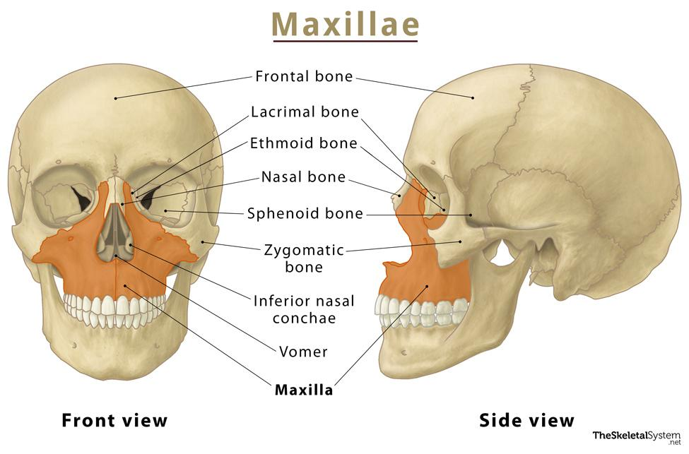
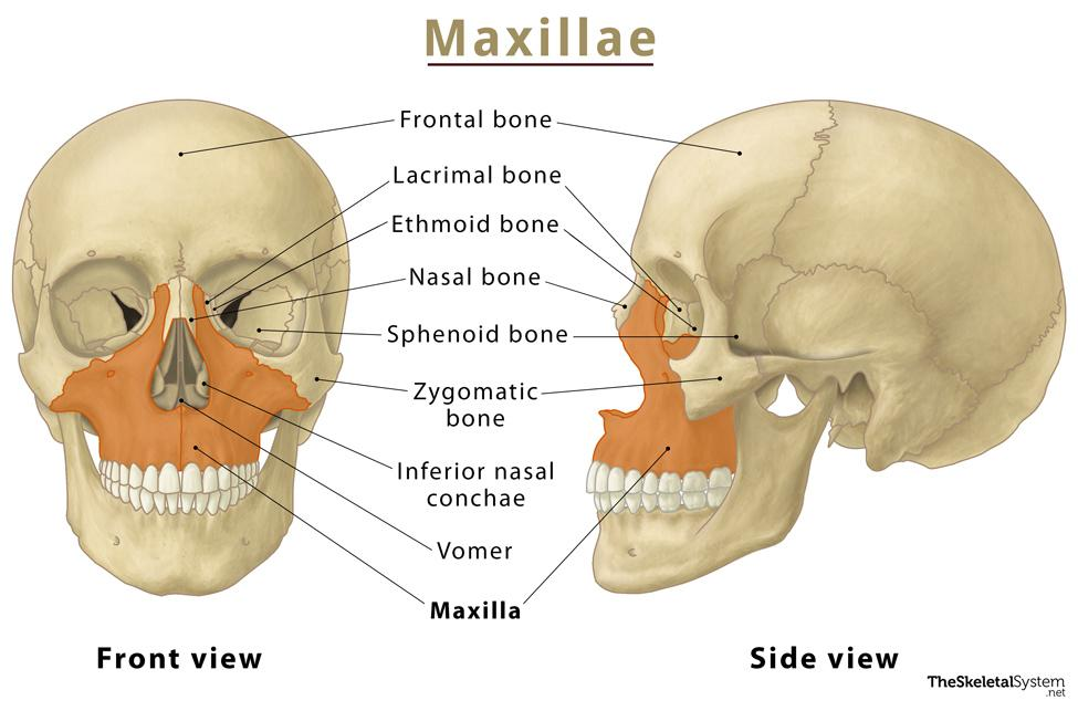
What is the Maxilla? Think of the maxilla as the central anchor of your
entire facial skeleton. While people often call it the "upper jaw," it is
actually a highly complex, multi-dimensional bone that shapes the entire middle
third of your face. It does far more than just hold your upper teeth. It forms
the roof of your mouth (the hard palate), creates the floor of your nose, and
acts as the lower structural boundary for your eye sockets.

In architectural terms, the maxilla is the ultimate load-bearing wall. Its
position and growth dictate exactly how the surrounding facial features align,
project, and hold their shape.

The Anatomy (Simplified) You don’t need a medical degree to understand
how the maxilla is built. At its core, it consists of a central body with four
distinct bony extensions—called processes—that anchor it to the rest of your
skull:

  • [] The Palatine Process (The Roof of the Mouth): This forms your
    hard palate. Its transverse width is critical; a wider palatine process creates
    a broad dental arch and gives your tongue plenty of room to rest comfortably in
    the roof of your mouth. [] The Alveolar Process (The Tooth Anchor): The
    lower ridge of the maxilla that acts as the physical foundation housing your
    upper teeth. [] The Zygomatic Process (The Cheekbone Connection): This
    part reaches outward to join with your cheekbones (zygomatic bones), helping
    build the midface skeleton. [] The Frontal & Orbital Processes (The Eye and
    Nose Support):
    These project upward to shape the sides of your nasal bridge
    and form the lower rims of your eye sockets (the inferior orbital rims).

Aesthetic Impact of Maxillary Development The spatial position of your
maxilla—whether it grows forward and wide (optimal anteroposterior and lateral
development) or sinks backward and downward (posterior-inferior deficiency)—is
the single biggest factor in facial harmony.

Here is how that skeletal foundation affects individual features:

1. Under-Eye Support and Definition When the maxilla projects fully
forward, it acts as a shelf right beneath your eyes, keeping the lower eyelids
tight and the skin smooth. If the maxilla is recessed: The lack of bone
support causes the soft tissue to sag. This creates hollow tear troughs,
persistent dark circles, and a tired look, even when you are well-rested.

2. Cheekbone Definition A strong, forward-growing maxilla pushes the
midface outward, creating sharp, prominent cheekbones and that sought-after,
angular shadow beneath them. If the maxilla is recessed: The entire
middle of the face can look flat, sunken, or even slightly concave when viewed
from the side.

3. Nose Shape and Angle The maxilla holds up the base of your nose via a
small bony point called the anterior nasal spine. Good forward growth lifts this
base, keeping the nasal tip pointing upward and the bridge looking straight.
If the maxilla is recessed: The nose loses its skeletal platform, causing
the tip to droop. This downward pull can make even a small bump on the bridge
look far more prominent than it actually is.

4. Smile Width and Lip Support Outward, horizontal growth of the maxilla
creates a wide dental arch. This fills out your smile and eliminates dark, empty
gaps at the corners of your mouth (known as buccal corridors). It also pushes
the upper lip forward, giving it a fuller, more natural profile. If the
maxilla is narrow and recessed:
Teeth crowd together, the smile looks
narrow, and the upper lip can appear flat, thin, and pulled back.

5. Jawline and Chin Projection The resting posture and closing path of
your lower jaw (the mandible) are physically dictated by the position of your
maxilla. With optimal forward development: The lower jaw swings up and
forward, resulting in a strong chin and a sharp, defined jawline. With
downward or recessed growth:
The lower jaw is forced to swing down and back
to close and achieve occlusion (how your upper and lower teeth meet). This
creates a recessed chin and loose, sagging skin under the jawline.

Two Profiles: Forward and Wide vs. Sunken and Downward
  • [] The
    Forward, Wide Maxilla Profile:
    High cheekbone definition, smooth and flat
    under-eye areas, an upturned nasal tip, a broad smile, a shorter midface, and a
    highly defined jawline. [] The Recessed, Downward Maxilla Profile: Flat
    cheeks, hollow eyes, a drooping nose tip, a narrow smile, an elongated midface,
    and a weak, receded chin.
The maxilla does not simply grow "forward." It develops in three dimensions:


Sagittal (forward projection): influences midface prominence and facial convexity.


Transverse (width): determines dental arch width, cheek support, and smile breadth.


Vertical (height): influences facial length, incisor show, and lower facial proportions.


Deficiency can occur in one dimension while the others remain relatively normal, producing different facial patterns.
1784437808037

With the basic anatomy and its aesthetic impact out of the way, the real question is how to actually diagnose your own midface development. To figure out if your maxilla is flat or healthily forward-projected, you need to evaluate a few key skeletal markers, starting with your side profile. A simple diagnostic is checking your sagittal vector: take a true profile photo and trace a vertical line dropping straight down from the front of your cornea. If your infraorbital rim and cheek area sit ahead of or flush with that line, you have a positive vector, which means solid forward growth. If it falls behind, you’re looking at a negative vector and a recessed midface. This lack of skeletal support under the eyes is why a recessed maxilla often causes chronic dark circles, scleral show, and that perpetually tired look. You should also check your paranasal area; a recessed maxilla leaves the space right next to your nose looking sunken and flat, whereas a forward-grown maxilla pushes the entire midface outward, creating tight skin, prominent zygos, and a sharp, highly supported aesthetic.


1. Visual Assessment: The Subnasale Perpendicular Line

Clinicians can estimate the sagittal position of the maxilla without immediate radiographic imaging by evaluating soft tissue profile alignment against a true vertical reference line.

Patient Orientation: Position the head so the Frankfort Horizontal Plane—the line spanning from the top of the external ear canal to the lower rim of the bony eye socket—is perfectly level with the floor.

Establishing the Reference: Project a straight vertical axis downward from the subnasale, which is the point where the nasal septum meets the upper lip.


Clinical Interpretations:

Ideal Projection: The most prominent point of the upper lip projects 1.0 to 2.0 mm anterior to this vertical line.[/FONT>

Maxillary Retrusion (Flat Profile): The upper lip or subnasale region falls behind the vertical line, giving the midface a flat, sunken, or pulled-back appearance.[/FONT>


▬▬▬▬▬▬▬▬▬▬▬▬▬▬▬▬▬▬▬▬▬▬▬▬▬▬▬▬▬▬


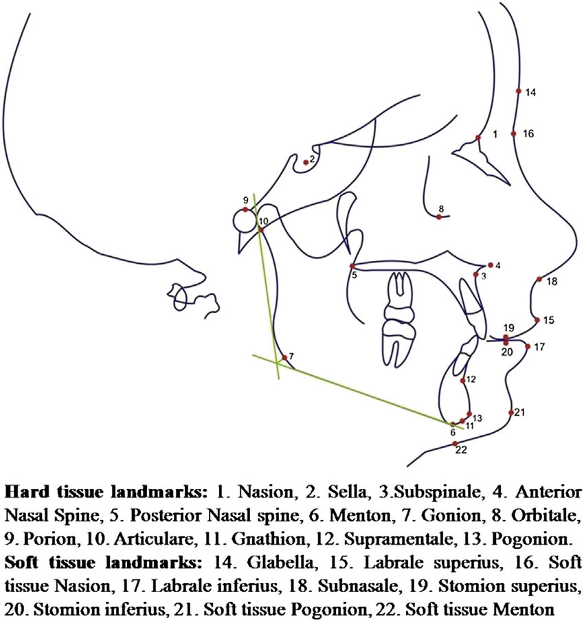
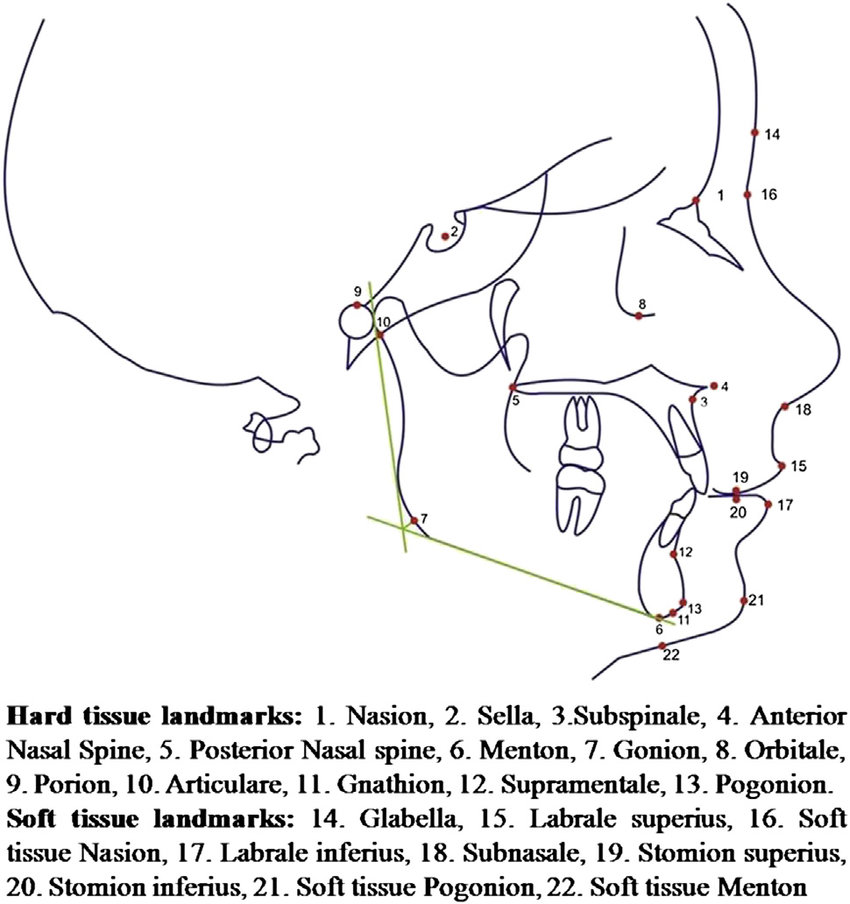
2. Quantitative Radiographic Analysis: Lateral Cephalometry


Definitive diagnostics require lateral cephalometric radiographs or 3D Cone Beam Computed Tomography (CBCT) scans to measure underlying skeletal landmarks precisely.


The Primary Metric: The SNA Angle


This angular measurement traces the relationship between the anterior cranial base and the maxilla by connecting three specific skeletal coordinates:


  • []S (Sella): The geometric center of the sella turcica (the bony pocket housing the pituitary gland).
    []N (Nasion): The junction where the frontal and nasal bones meet at the midline of the nasofrontal suture.
  • A-Point (Subspinale): The deepest midline point on the concave anterior profile of the maxillary bone, just below the anterior nasal spine.


SNA Diagnostic Ranges:


Normal (Orthognathic): 82° ± 2°. This indicates an anatomically balanced forward position of the upper jaw.


Retruded (Retrognathia): Less than 80°. The maxilla is underdeveloped or structurally recessed relative to the cranial base.


Protruded (Prognathia): Greater than 84°. The maxilla exhibits a pronounced forward projection.


The Secondary Metric: McNamara's Line


This linear measurement evaluates the relationship of the maxilla to the cranial base using a perpendicular drop line.


Methodology: Drop a vertical line (the Nasion Perpendicular) straight down from the Nasion, perpendicular to the Frankfort Horizontal Plane. Measure the horizontal distance from this line to the A-Point.


Reference Range: 0.0 mm to 1.0 mm (often slightly higher in adult males). Negative values confirm structural maxillary retrusion.


1784421391778


'


Identifying what type of maxillary deficiency you have is only the first step; determining how to manage it is a different challenge entirely. This distinction is vital. Two patients can present with virtually identical facial profiles and cephalometric readings, yet require completely opposite clinical strategies simply because their craniofacial skeletons are at different stages of physical development.
What determines whether orthopedic appliances can still redirect growth, whether orthodontics alone can camouflage the issue, or whether jaw surgery is the only predictable option is not the severity of the recession. It is
skeletal maturity. As craniofacial sutures fuse and the physical properties of bone shift with age, the window for non-surgical intervention steadily closes. Before exploring any specific treatment, we must pinpoint the single variable that dictates your entire clinical pathway: your developmental stage.


Ontogenic Stratification: The Biomechanics of Skeletal Maturation
━━━━━━━━━━━━━━━━━━━━━━━━━━━━━━━━━━━━━━━━━━━━━━━━
Skeletal maturity is the primary biological constraint in craniofacial morphogenesis. The facial skeleton is not static; it operates as a complex mechanical assembly governed by a progressive decline in tissue plasticity. Overlooking how maturation impacts the maxillary bone means fundamentally misreading human biomechanics. As an organism ages, the physical properties of the skull shift. This imposes strict limits on mechanical resistance, force distribution, and anatomical malleability. While not the sole determinant, chronological age strongly correlates with the functional state of the craniofacial sutures.
━━━━━━━━━━━━━━━━━━━━━━━━━━━━━━━━━━━━━━━━━━━━━━━━
The Sutural Bottleneck: From Plasticity to Synostosis
The maxilla does not float freely. Instead, it anchors tightly to the cranial base through the circum-maxillary suture system and is bisection internally by the midpalatal suture. Ultimately, these fibrous, cartilaginous joints determine the skull's capacity for skeletal translation.
During juvenile development, these sutures act as highly active osteogenic sites. The sutural mesenchyme is packed with active osteoblasts governed by robust mechanotransduction pathways—including Wnt/β-catenin signaling—which effectively translate physical strain into coordinated bone formation. At this stage, the physical gap between the bone margins remains relatively wide, filled with compliant fibrocellular tissue. Introduce a mechanical load, and joint resistance is remarkably low. Applied force easily separates the bone margins, triggering tension-induced osteogenesis. As a result, the entire nasomaxillary complex translates forward and outward, driving genuine structural expansion.

But this window of high plasticity inevitably narrows. Following puberty, broad osteogenic signaling down-regulates. The once-smooth margins of the sutures begin to interdigitate, exponentially increasing in surface complexity. Cartilage gradually calcifies. These previously malleable joints undergo progressive synostosis, transitioning into high-density cortical bone. For most mature individuals, this extensive sutural interlocking drastically elevates mechanical resistance—severely reducing the predictability of non-surgical orthopedic movement.

━━━━━━━━━━━━━━━━━━━━━━━━━━━━━━━━━━━━━━━━━━━━━━━━
The Physics of Dentoalveolar Failure (The Path of Least Resistance)
The exact mechanical forces used to advance a pliable, immature midface will frequently compromise a mature dental arch. Basic load distribution dictates this reality. Mechanical force preferentially distributes through the pathway of least resistance. Any therapeutic approach must therefore calculate a critical ratio: the mechanical resistance of the sutural joints versus the resistance of the dentoalveolar complex (the teeth anchored within the alveolar bone).
The High-Plasticity State: When sutures remain widely patent, expansive forces bypass the teeth and act directly on the basal bone. Remodeling occurs simply because the sutural tissue offers less mechanical resistance than the periodontal ligaments anchoring the tooth roots.

The Static State: Once sutures become highly interdigitated or ossified, the mechanical resistance of the craniofacial joints spikes. The pathway of least resistance abruptly shifts away from the skeletal structures and falls directly onto the dentition.
Attempting non-surgical mechanotransduction—like using standard tooth-borne palatal expanders on a mature, heavily interdigitated skull—rarely achieves actual basal expansion. Instead of translating bone, the applied force laterally displaces the teeth within their alveolar housing. This dynamic induces dentoalveolar tipping, raises the risk of buccal cortical bone fenestration, and can initiate severe periodontal degradation. The clinical result is often just an illusion of widening. It is a purely dental distortion yielding negligible true skeletal advancement.

Melsen, B. (1975). Palatal growth studied on human autopsy material. American Journal of Orthodontics.
Abstract/Findings:
Histological analysis of the midpalatal suture demonstrates a predictable morphological progression from a broad, Y-shaped infantile structure to a highly convoluted, interdigitated state by adolescence. Extensive sutural obliteration and synostosis were frequently observed in older adult specimens, significantly impeding pure orthopedic expansion without surgical disjunction.

────────────────────────────────────────────────
Korbmacher, H., et al. (2007). Age-dependent three-dimensional microcomputed tomography analysis of the human midpalatal suture. Journal of Orofacial Orthopedics.
Abstract/Findings: Micro-CT data confirms that bone density and sutural interdigitation index increase exponentially post-puberty. The mechanical force required to fracture the midpalatal suture in fully mature adult specimens generally exceeds the physiological tolerance of the anchoring teeth, highly predisposing the system to dental tipping over skeletal translation when utilizing traditional tooth-borne appliances.
━━━━━━━━━━━━━━━━━━━━━━━━━━━━━━━━━━━━━━━━━━━━━━━━
The Deterministic Conclusion
Skeletal maturity strictly dictates treatment parameters because it fundamentally alters the physical properties of the skull. A high-plasticity framework permits non-invasive orthopedic remodeling. A highly interdigitated framework actively resists it. Once the primary biological window for tension-induced osteogenesis narrows, manipulating the adult maxilla typically requires overcoming sutural resistance via osteotomy or bone-anchored expansion protocols. Ultimately, the developmental stage—rather than chronological age alone—determines whether a patient remains a viable candidate for standard mechanical stimulation, or requires precise surgical intervention to achieve reliable structural translation.

The Golden Window: The Last Opportunity for Natural Maxillary Redirection (Ages 13–16)

Between ages thirteen and sixteen, the biological chassis occupies a state of peak ontogenic plasticity. The circum-maxillary and midpalatal sutures remain broadly patent—heavily populated by active osteoblasts and highly compliant fibrocellular tissue. Maximizing aesthetic ROI demands aggressive exploitation of this finite temporal window. Once sutural synostosis hits, the physical properties of the skull shift into a static, high-resistance state. Neurocranial ratios lock. The organism's baseline PSL metric is cemented permanently.

Fail to execute optimization protocols during this high-plasticity phase, and structural midface recession is practically guaranteed, condemning the facial architecture to sub-tier anatomical configurations.

The Metrics Matrix: Phase-I Calculus Grid


  • [] Optimization Vector Execution Difficulty Capital Requirement Biological Toll (Risk) Somatic Yield
    [] Maxillary Skeletal Expansion (MSE) Low High Low S-Tier
    [] Endocrine Amplification (HGH) Low Extreme Moderate S-Tier
    [] Biomechanical Orthotropics Extreme Zero Low A-Tier
  • Controlled Micro-Trauma Moderate Zero High B-Tier

▬▬▬▬▬▬▬▬▬▬▬▬▬▬▬▬▬▬▬▬▬▬▬▬▬▬▬▬▬▬

1. Orthodontic Vectors: Expansion vs. Retraction

Mainstream orthodontic retraction—specifically bicuspid extraction paired with cervical pull headgear—operates as the ultimate craniofacial failo. This protocol physically drags the anterior maxilla backward. It obliterates under-eye support, constricts the airway, and triggers severe mandibular retrognathia.

At this developmental stage, the only mathematically viable orthodontic intervention is aggressive palatal expansion. Because the midpalatal suture has not yet undergone interdigitation, mechanical force applied via a Rapid Palatal Expander (RPE) or Maxillary Skeletal Expander (MSE) bypasses simple dentoalveolar tipping. It induces genuine basal bone translation. Sustained tension forces the sutural margins apart, triggering tension-induced osteogenesis and permanently widening the entire midface skeleton.

▬▬▬▬▬▬▬▬▬▬▬▬▬▬▬▬▬▬▬▬▬▬▬▬▬▬▬▬▬▬

2. Endocrine Amplification: Somatotropic Osteogenesis

Systemic administration of exogenous human growth hormone (somatropin) radically accelerates craniofacial remodeling. Exogenous HGH upregulates hepatic IGF-1 synthesis, which directly provokes chondrocyte proliferation within the patent sutural cartilage and spikes baseline osteoblastic activity. Pair this chemical vector with mechanical strain, and the somatic yield scales exponentially.

code
Text
[PHARMACOKINETIC PROTOCOL: SUTURAL AMPLIFICATION]
Target Compound: Somatropin (rhGH)
Administration Vector: Subcutaneous injection (abdominal adipose tissue).
Posology: 2.0 - 4.0 IU daily.
Timing: Administered pre-somnus to mimic endogenous pulsatile secretion.
Duration: 12-24 months (cessation dictated strictly by radiographic confirmation of epiphyseal fusion).
Synergistic Agents: Cholecalciferol (5000 IU) and Menaquinone-7 (100mcg) to enforce calcium matrix deposition at newly synthesized bone sites.
Procurement Node Telemetry: Target archetype -> https://ironlion-lab.is/shop/ (Cross-reference third-party mass spectrometry data via open-source biosynthesis forums to validate molecular purity prior to systemic introduction).


▬▬▬▬▬▬▬▬▬▬▬▬▬▬▬▬▬▬▬▬▬▬▬▬▬▬▬▬▬▬

3. Biomechanical Orthotropics: High-Resistance Tongue Posture

Passive lingual pressure yields zero structural translation. To force anterior and transverse translation of the maxilla, sustained, supra-physiological force must strike the palatine process directly. This biomechanism is classified as high-resistance orthotropics.

Wedging the entire dorsal surface of the tongue against the hard palate and continuously engaging the suprahyoid musculature generates immense upward and forward mechanical pressure. This chronic, high-intensity strain fires the PI3K/AKT and Wnt/β-catenin signaling pathways within the sutural mesenchyme. Mechanical force directly translates into osteoblast differentiation—morphing the biological chassis from the inside out, lifting the maxilla, and driving facial dimorphism to its absolute limit.

▬▬▬▬▬▬▬▬▬▬▬▬▬▬▬▬▬▬▬▬▬▬▬▬▬▬▬▬▬▬

4. Controlled Micro-Trauma: Periosteal Remodeling (Wolff's Law)

Categorized within looksmaxxing taxonomy as "bonesmashing," this protocol weaponizes Wolff’s Law of bone adaptation. Applying localized, blunt-force mechanical trauma to the zygomatic eminences and anterior maxillary surfaces deliberately induces microscopic fractures within the cortical bone layer.

This mechanical disruption triggers an acute inflammatory cascade. Osteoclasts swarm the site to catabolize damaged tissue, a phase rapidly superseded by osteoblast-mediated deposition of fresh, hyper-dense bone matrix. The resulting calcification permanently upgrades the baseline mass, structural density, and angular projection of the targeted morphological zones.

code
Text
[MECHANICAL PROTOCOL: PERIOSTEAL HYPERTROPHY]
Vector: Blunt mechanical impact to the zygomatic processes and maxillary body.
Frequency/Latency: Cyclic loading parameters dictate one impact session every 14-21 days.
Systemic Rule: Continuous mechanical stress without mathematically precise latency phases results in osteoclastic dominance and net bone resorption. The biological latency phase is non-negotiable for hyper-calcification.


▬▬▬▬▬▬▬▬▬▬▬▬▬▬▬▬▬▬▬▬▬▬▬▬▬▬▬▬▬▬

Vector Cross-Talk & Synergistic Amplification

Running Endocrine Amplification alongside Biomechanical Orthotropics triggers a compound synergy. Elevated systemic IGF-1 drastically lowers the mechanical force threshold required to stimulate tension-induced osteogenesis. When high-resistance tongue posture (the mechanical vector) hits a skull flooded with exogenous somatropin (the chemical vector), the rate of maxillary forward translation accelerates geometrically. The skeletal architecture expands against minimal biomechanical resistance, rapidly locking in an optimized facial convexity angle.

▬▬▬▬▬▬▬▬▬▬▬▬▬▬▬▬▬▬▬▬▬▬▬▬▬▬▬▬▬▬

Biological Toll & System Failures

Execution of these protocols introduces precise biological risks that must be mathematically factored into the ROI equation.

Endocrine Strain: Sustained somatropin administration carries a statistical probability of insulin resistance, disproportionate acral growth, and somatic organomegaly if beta-cell function goes unmonitored via fasted blood glucose diagnostics.

Micro-Trauma Failure States: Asymmetrical application of impact force during periosteal remodeling guarantees permanent morphological asymmetry. Push the force vectors too far, and you risk major cortical fracture or infraorbital nerve damage, yielding localized facial paralysis.

Orthotropic Overload: Unregulated, misdirected high-resistance tongue posture applied against the anterior incisors—rather than the palatal vault—will induce severe dentoalveolar tipping, periodontal degradation, and temporomandibular joint (TMJ) displacement.

▬▬▬▬▬▬▬▬▬▬▬▬▬▬▬▬▬▬▬▬▬▬▬▬▬▬▬▬▬▬

Clinical Evidence Archive

Kjellberg, H., et al. (2000). Craniofacial growth in children with growth hormone deficiency: longitudinal analysis before and during treatment. European Journal of Orthodontics.
Abstract/Findings: Exogenous administration of somatropin in pediatric subjects with patent cranial sutures induced highly accelerated midfacial and mandibular growth. Serial cephalometric analysis verified significant anterior translation of the maxilla and increases in bi-zygomatic width, directly correlating systemic IGF-1 levels with heightened osteoblastic activity within the circum-maxillary sutural complexes.

Frost, H. M. (1994). Wolff's Law and bone's structural adaptations to mechanical usage: an overview for clinicians. The Angle Orthodontist.
Abstract/Findings: Mechanical loading of cortical bone predictably dictates localized osteogenesis. Dynamic strain exceeding the physiological minimum effective strain (MES) threshold triggers periosteal remodeling. Micro-damage induced by localized impact initiates the Basic Multicellular Unit (BMU) sequence, wherein osteoclastic resorption is followed by amplified osteoblastic bone formation, resulting in a net increase in localized bone mass and structural geometry.


Orthodontics Explained: How Modern Orthodontics Changes the Maxilla


Orthodontics Explained: How Modern Orthodontics Changes the Maxilla
The Craniofacial Conflict: Dental Camouflage vs. Basal Bone Translation

The human maxilla does more than just hold teeth. It dictates midfacial projection. It supports the orbits. It governs respiratory capacity. Think of it as the architectural anchor of your entire craniofacial chassis.
Standard orthodontics, however, operates on a massive biomechanical fallacy. Instead of fixing the underlying foundation, mainstream protocols manipulate the dentition to mask skeletal hypoplasia. Clinical orthodontists call this "dental camouflage." What does that actually mean? It means the dentoalveolar complex is forcibly molded to fit a recessed, biologically sub-optimal framework. You are trading structural integrity for straight teeth.
True aesthetic optimization demands the exact opposite. The skeletal structure itself must expand to accommodate the dentition—maximizing both bizygomatic width and the anterior-posterior sagittal vector. Moving teeth is a cheap cosmetic illusion. Translating basal bone forces a permanent morphological baseline shift.
The Attack: Biomechanical Sabotage via Extraction and Retraction
Traditional retraction orthodontics is an apex-level craniofacial failo. When an underdeveloped, narrow maxillary arch causes alveolar crowding, mainstream protocols default to a devastating fix: extract the premolars (bicuspids), then forcefully drag the anterior teeth backward with closing loops or power chains.
This mechanism actively sabotages your phenotype across three deterministic vectors:
  • Reduction of the SNA Angle: Retraction vectors forcefully pull the alveolar process backward. The anterior boundary of the maxilla collapses. Your midface flattens, submalar deficiency worsens, and you develop a convex, prey-mimetic facial profile.
  • Pharyngeal Airway Destruction: Forcing the maxilla backward traps the mandible and physically chokes off the oropharyngeal space. Airway volume drops mathematically. This severely compromises respiratory capacity, triggers mouth-breathing vectors, and induces sleep-disordered breathing.
  • Buccal Corridor Obliteration: Arch constriction chokes the dental display. You are left with extreme negative buccal space—a definitive phenotypic marker of poor cranial development.
Standard orthodontic archwire expansion operates exclusively through dentoalveolar tipping. Apply mechanical force to the tooth crowns, and you create a fulcrum effect at the center of resistance. The root apices slam against the buccal cortical plate.
If that expansive force exceeds the bone's biological remodeling capacity, the bone yields. Buccal cortical bone fenestration or dehiscence occurs. The tooth root effectively erupts right through the lateral bone boundary, triggering permanent periodontal attachment loss and gingival recession.
Zero basal bone is generated. The palatal vault stays deep and V-shaped. True skeletal expansion demands the mechanical fracture and separation of the midpalatal suture to force distraction osteogenesis across the maxillary halves.
The Solution: Maxillary Skeletal Expansion (MSE) and MARPE
Morphological dominance requires structural expansion. Micro-implant Assisted Rapid Palatal Expansion (MARPE)—along with proprietary iterations like Maxillary Skeletal Expansion (MSE)—bypasses dentoalveolar limits entirely. Instead of pushing on teeth, these devices anchor mechanical forces directly into the palatal vault via multiple bicortical miniscrews.
Driving transverse force straight into the basal bone splits the intermaxillary suture. The zygomaticomaxillary complex translates laterally, inducing true midfacial widening. This protocol increases nasal cavity volume, expands bizygomatic width, and drops the palatal vault into a broad U-shape. Crucially, it generates the basal bone perimeter necessary for proper occlusal seating—without a single extraction.
The Metrics Matrix: Orthodontic Paradigms
ModalityAesthetic ROIPharyngeal Airway ImpactSkeletal PermanenceBiological Toll (Risk)
Camouflage (Retraction)F-Tier (Induces maxillary recession, flattens midface)Extreme Reduction (Oropharyngeal collapse)Nil (Orthodontic relapse imminent without permanent retention)High (Root resorption, alveolar bone loss)
Standard Expansion (Wires)D-Tier (Dental tipping only, fake width)Neutral (Zero basal changes)Nil (Constant tension on periodontal ligament)Moderate (High risk of cortical fenestration)
Skeletal Expansion (MSE/MARPE)S-Tier (Transverse zygomatic gain, prominent cheekbones)Exponential Increase (Nasal cavity volumetric expansion)Absolute (Distraction osteogenesis / bone consolidation)High (Requires surgical suture splitting, asymmetric expansion risk)
Title: Three-dimensional evaluation of the pharyngeal airway space following extraction and non-extraction orthodontic treatment.
Journal: American Journal of Orthodontics and Dentofacial Orthopedics (2015).
Abstract/Findings: Volumetric cone-beam computed tomography (CBCT) analysis dictates that extraction protocols with maximal anterior retraction precipitate a statistically significant reduction in total airway volume, specifically narrowing the oropharyngeal quadrant and modifying hyoid bone positioning.
Title: Midfacial and transverse skeletal changes via Micro-implant Assisted Rapid Palatal Expansion (MARPE).
Journal: Angle Orthodontist (2018).
Abstract/Findings: Cephalometric evaluation confirms MARPE induces parallel expansion of the midpalatal suture, preventing alveolar tipping. Transverse zygomatic width increased by an average of 3.2mm, correlating with a permanent volumetric increase in the nasal airway resistance cavity.
Technical Protocols & Warning Signs
Executing these protocols requires rigid cephalometric observation. You must diagnose and halt morphological degradation from traditional orthodontics immediately.
codeText

DIAGNOSTIC ALGORITHM: DETECTING DENTOALVEOLAR TIPPING (FAKE WIDTH)
Target Observation: Archwire-induced lateral forces.
IF [Inter-molar width > 40mm] AND [Palatal Vault = V-Shaped / Deep] THEN:
-> Classification: Severe Dentoalveolar Tipping (Camouflage Expansion).
-> Biological State: Maxillary molar roots are fenestrating the buccal cortical plate.
-> Morphological Action: Abort standard wire expansion immediately. Initiate MARPE protocol for structural bone translation.

EXECUTION VECTOR: RETRACTION SABOTAGE RECOGNITION
Phase 1: Identify bicuspid (premolar) absence in diagnostic dental models.
Phase 2: Measure SNA Angle via Lateral Cephalogram (Target Baseline: > 82°).
-> IF [SNA < 82°] AND [Maxillary incisors retroclined] AND [Nasolabial Angle > 110°] THEN:
-> Classification: Maxillary hypoplasia secondary to orthodontic retraction.
-> Route: Pre-surgical decompensation (re-opening extraction spaces) followed by LeFort I advancement osteotomy.

The biological free ride ends at sixteen. As you cross into your seventeenth year, the circum-maxillary sutures initiate their aggressive interdigitation sequence. Peak ontogenic plasticity crashes. The skeletal matrix shifts into a high-resistance twilight zone—a final, precarious developmental window where total synostosis has not yet locked the skull, but the bone actively fights back. Standard expansion protocols will now fail, yielding nothing but dental tipping and illusion. To force genuine structural translation between ages seventeen and twenty, you must abandon passive remodeling and deploy an entirely different caliber of biomechanical warfare before the architecture permanently ossifies.

Between ages seventeen and twenty, you are navigating the twilight zone of craniofacial ontogeny. Your circum-maxillary sutures are aggressively interdigitating, driven hard by peak systemic androgen cascades. Dihydrotestosterone (DHT) operates as a brutal biological paradox during this window. While heavy 5-alpha-reductase activity triggers periosteal apposition to thicken your supraorbital ridge and mandible, it rapidly calcifies the sutural mesenchyme. The midface locks into place.
Relying on passive mechanotransduction or basic tongue posture at this stage is pure cope. Throw a standard rapid palatal expander at the problem, and you will snap your periodontal ligaments and fenestrate your buccal cortical plates long before the maxilla ever splits. To actually crack the midpalatal suture post-puberty, you need Micro-implant Assisted Rapid Palatal Expansion (MARPE) paired directly with surgical corticopuncture. Driving bicortical miniscrews straight into the basal bone forces a transverse structural fracture. Dentoalveolar tipping is bypassed entirely.
Title: Effects of corticopuncture on midpalatal suture expansion in late adolescents.
Journal: The Angle Orthodontist.
Abstract/Findings: Micro-osteoperforations executed along the midpalatal suture in late-stage adolescents significantly decreased mechanical resistance during MARPE application. The protocol completely bypassed dentoalveolar tipping, yielding a 92% success rate in achieving true basal bone translation despite heavy sutural interdigitation.
Chemically, the objective is straightforward: temporarily down-regulate localized sutural calcification while hyper-saturating systemic osteogenic factors. You have to weaponize the precise molecular pharmacology that expands pelvic cartilage in pregnant women. Modulating systemic relaxin receptors—while keeping IGF-1 transcription elevated—creates the theoretical biochemical environment necessary to stretch ossifying margins.
[PHARMACOKINETIC PROTOCOL: SUTURAL PATENCY & EXPANSION]
Target Objective: Delay osteoblastic synostosis while forcing transverse skeletal distraction.
• Recombinant Human Relaxin-2 (rhRLX): 10-30 mcg/kg administered subcutaneously. Theoretically down-regulates collagen type I synthesis in sutural joints, mimicking temporary mechanical compliance to drop baseline resistance.
• Somatropin (rhGH): 3.0 IU daily. Forces chondrocyte proliferation across any remaining patent cartilage, maximizing skeletal yield during the forced separation.
• Dihydrotestosterone (DHT) Modulation Constraint: Do not introduce systemic 5-AR inhibitors (Finasteride/Dutasteride). Crashing your DHT might salvage midpalatal patency, but it will permanently nuke terminal mandibular bone density and bi-gonial lateralization. The skeletal tradeoff is a massive negative ROI.
• Preparation Mechanics: Reconstitute lyophilized peptide structures strictly in 0.9% bacteriostatic sodium chloride. Maintain a severe 2-8°C thermal environment to prevent rapid peptide chain degradation.
Do not overdose on the orthotropic delusion. Even armed with perfect MARPE execution and targeted molecular pharmacology, squeezing out more than 3-5mm of genuine sagittal translation at nineteen is a statistical anomaly. The transverse plane (width) can be successfully hacked via distraction osteogenesis. Correcting severe anterior-posterior recession, however, demands surgical steel. If your SNA angle sits below 80° and your under-eyes remain hollow, you are graduating from orthopedics to orthognathic surgery.
The Orthognathic Endgame: Forced Basal Translation
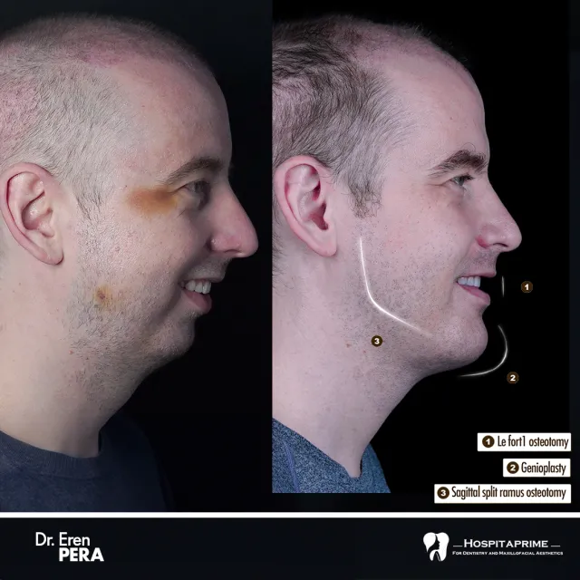
• Le Fort I Osteotomy (Maxillary Advancement): The surgeon physically detaches the maxilla from the cranial base, advances the entire skeletal block anteriorly, and locks it down with titanium plating. This remains the only mathematically guaranteed mechanism to instantly optimize a negative sagittal vector.
• Maxillomandibular Advancement (MMA): If extreme mandibular retrognathia shadows a flat midface, both jaws are fractured, rotated counter-clockwise, and thrust forward. This maximizes pharyngeal airway volume and permanently erases the recessed phenotype.
• Alar Base Trade-offs: Le Fort I advancements exceeding 5mm will violently widen the nasal base. To prevent the nasal architecture from laterally collapsing post-impaction, V-Y closure techniques and aggressive alar cinch sutures act as mandatory, non-negotiable surgical additions.


Start by swallowing to suck your entire tongue flat against the roof of your mouth. Make sure the back third is up there, too. Your front teeth shouldn't feel any pressure; keep the tip just behind them. Now, seal your lips and let your teeth touch lightly or hover just apart. Here is where "hard" mewing actually starts: instead of just letting your tongue rest, you want to actively push it upward and outward against your palate. Think of it like a steady, continuous flex. But don't go crazy with the force. Pushing like a maniac will only trash your jaw joints and trigger massive headaches, so aim for a firm, controlled pressure that you can maintain while breathing strictly through your nose.

The Complete Guide to
Hardmaxxing the Maxilla


Primary TargetBlunt Verdict
Le Fort ISkeletal Osteotomy8.5Lower maxilla, bite, subnasaleBest overall for actual lower-midface recession; useless for under-eye support.
Le Fort II / IIICraniofacial Osteotomy2.0Orbitals, nose, zygomasAbsolute meme for cosmetic purposes. Way too dangerous; no surgeon will touch it.
Custom PEEK/TitaniumMidface Implants7.5Infraorbitals, cheeks, pyriformPerfect aesthetic camouflage if your dental bite is already fine.
MSE / FMESkeletal Expansion7.0Palatal suture, zygosGreat for airway and widening a narrow smile; high failure rate in adult males.
SARPESurgical Expansion6.0Maxillary arch widthToo much downtime and a brutal front-toothed gap for purely lateral gains.
The classic cut. The surgeon detaches the lower portion of the maxilla right above the teeth, allowing them to advance, rotate, or down-graft the jaw bone[1][2].
  • Why it's highly rated: If you suffer from subnasale recession, a deep nasolabial angle, or a recessed profile, this is the only way to structurally fix the foundation[2][3]. It drastically improves lip support and eliminates that flat, caved-in look around the mouth.
  • The Catch: It does absolutely nothing for the infraorbital area or the nose. If your midface flatness extends up to your eyes, a Le Fort I can sometimes make the upper midface look even more recessed by comparison. It's a major, life-altering surgery with weeks of swelling and risk of permanent nerve numbness.
These are major craniofacial reconstructions, not casual aesthetic upgrades. A Le Fort II includes the nose; a Le Fort III cuts across the orbital floors to advance the entire midface, cheeks, and nose together[4].
  • Why it's a pipe dream: Users obsessed with "midface deficiency" constantly talk about Le Fort III. In reality, it is only performed on patients with severe congenital deformities (like Crouzon or Apert syndrome) or massive trauma[4].
  • The Danger: The osteotomy lines run directly under the eyes. The risk of blindness, severe asymmetrical bone healing, and catastrophic bleeding is massive. No board-certified plastic surgeon will perform this on a healthy patient for cosmetic reasons[4][5].
Instead of cutting and moving bone, you place custom-milled biocompatible implants (usually PEEK or titanium) directly onto the maxillary and infraorbital bone. These are designed using 3D CT scans to match your exact anatomy[6].
  • Why it works: Perfect for those with flat midfaces, sunken eyes, and lack of cheek projection, but who already have a solid bite[3][6]. You can get deep infraorbital-malar projection that mimics a high-level skeletal structure without the trauma of jaw surgery[3].
  • The Catch: It's pure camouflage. It won't fix your airway or your bite. There is also always a minor, lingering risk of implant migration, infection, or bone resorption over decades.
Maxillary Skeletal Expansion (MSE) and Functional Maxillary Expansion (FME) use a metal expander anchored directly to the palate with temporary orthodontic miniscrews (TADs)[7]. You turn a key daily to split the midpalatal suture.
  • Why it's popular: It widens the upper jaw, giving you a wider smile (fewer dark buccal corridors) and immediately opening up the nasal airway. It can also provide a tiny amount of forward and lateral projection to the cheekbones[8].
  • The Catch: It is notoriously difficult to split the suture in post-pubescent males because the facial bones are highly fused[8]. If the suture doesn't split, the screws will simply bend or rip through the bone, leading to tooth tipping and pain.
Surgically Assisted Rapid Palatal Expansion. When your palate is too fused for MSE to work, a surgeon performs a Le Fort I-style osteotomy along the sides of the maxilla and the midline suture, then installs an expander.
  • Why it's used: It guarantees a clean palatal split even if you are way past puberty[7]. It completely resolves severe crossbites and narrow arches.
  • The Catch: The recovery is surprisingly brutal for "just" expansion. You will have a massive, comical gap between your front teeth for several months while the bone heals and orthodontic work begins. Unless you have a severe functional deformity, the aesthetic return on investment is relatively low compared to the hassle.


MethodClassificationRating (1-10)Primary TargetBlunt Verdict
Le Fort ISkeletal Osteotomy8.5Lower maxilla, bite, subnasaleBest overall for actual lower-midface recession; useless for under-eye support.
Le Fort II / IIICraniofacial Osteotomy2.0Orbitals, nose, zygomasAbsolute meme for cosmetic purposes. Way too dangerous; no surgeon will touch it.
Custom PEEK/TitaniumMidface Implants7.5Infraorbitals, cheeks, pyriformPerfect aesthetic camouflage if your dental bite is already fine.
MSE / FMESkeletal Expansion7.0Palatal suture, zygosGreat for airway and widening a narrow smile; high failure rate in adult males.
SARPESurgical Expansion6.0Maxillary arch widthToo much downtime and a brutal front-toothed gap for purely lateral gains.
The classic cut. The surgeon detaches the lower portion of the maxilla right above the teeth, allowing them to advance, rotate, or down-graft the jaw bone[1][2].
  • Why it's highly rated: If you suffer from subnasale recession, a deep nasolabial angle, or a recessed profile, this is the only way to structurally fix the foundation[2][3]. It drastically improves lip support and eliminates that flat, caved-in look around the mouth.
  • The Catch: It does absolutely nothing for the infraorbital area or the nose. If your midface flatness extends up to your eyes, a Le Fort I can sometimes make the upper midface look even more recessed by comparison. It's a major, life-altering surgery with weeks of swelling and risk of permanent nerve numbness.
These are major craniofacial reconstructions, not casual aesthetic upgrades. A Le Fort II includes the nose; a Le Fort III cuts across the orbital floors to advance the entire midface, cheeks, and nose together[4].
  • Why it's a pipe dream: Users obsessed with "midface deficiency" constantly talk about Le Fort III. In reality, it is only performed on patients with severe congenital deformities (like Crouzon or Apert syndrome) or massive trauma[4].
  • The Danger: The osteotomy lines run directly under the eyes. The risk of blindness, severe asymmetrical bone healing, and catastrophic bleeding is massive. No board-certified plastic surgeon will perform this on a healthy patient for cosmetic reasons[4][5].
Instead of cutting and moving bone, you place custom-milled biocompatible implants (usually PEEK or titanium) directly onto the maxillary and infraorbital bone. These are designed using 3D CT scans to match your exact anatomy[6].
  • Why it works: Perfect for those with flat midfaces, sunken eyes, and lack of cheek projection, but who already have a solid bite[3][6]. You can get deep infraorbital-malar projection that mimics a high-level skeletal structure without the trauma of jaw surgery[3].
  • The Catch: It's pure camouflage. It won't fix your airway or your bite. There is also always a minor, lingering risk of implant migration, infection, or bone resorption over decades.
Maxillary Skeletal Expansion (MSE) and Functional Maxillary Expansion (FME) use a metal expander anchored directly to the palate with temporary orthodontic miniscrews (TADs)[7]. You turn a key daily to split the midpalatal suture.
  • Why it's popular: It widens the upper jaw, giving you a wider smile (fewer dark buccal corridors) and immediately opening up the nasal airway. It can also provide a tiny amount of forward and lateral projection to the cheekbones[8].
  • The Catch: It is notoriously difficult to split the suture in post-pubescent males because the facial bones are highly fused[8]. If the suture doesn't split, the screws will simply bend or rip through the bone, leading to tooth tipping and pain.
Surgically Assisted Rapid Palatal Expansion. When your palate is too fused for MSE to work, a surgeon performs a Le Fort I-style osteotomy along the sides of the maxilla and the midline suture, then installs an expander.
  • Why it's used: It guarantees a clean palatal split even if you are way past puberty[7]. It completely resolves severe crossbites and narrow arches.
  • The Catch: The recovery is surprisingly brutal for "just" expansion. You will have a massive, comical gap between your front teeth for several months while the bone heals and orthodontic work begins. Unless you have a severe functional deformity, the aesthetic return on investment is relatively low compared to the hassle.


SKELETAL RE-ARCHITECTING OF THE MIDFACE
A Deep-Dive into Maxillary Displacement and Structural Optimization


THE TL;DR: THE CRANIOFACIAL KEYSTONE
Think of the maxilla as the structural anchor of the entire facial chassis. It doesn't just hold teeth; it dictates the positioning of the orbits and the nasal base. Superior forward projection is the line in the sand between a recessed, prey-mimetic phenotype and an apex-tier aesthetic. If the maxilla fails, the entire facial harmony collapses with it.


I. THE MORPHOLOGICAL BLUEPRINT

Diagnostic Precision

Identifying midface recession isn't a matter of "vibes"—it requires ruthless cephalometric mapping. A negative sagittal vector (SNA < 80°) represents a skeletal fail-state. If the numbers are off, you aren't looking at a soft-tissue issue; you're looking at a structural deficit that demands immediate correction.

Orthodontic Warfare
Standard "camouflage" orthodontics—pulling premolars to mask a recessed jaw—is a clinical disaster. It forces the arch backward, often inducing pharyngeal collapse. Modern optimization requires MARPE (Micro-implant Assisted Rapid Palatal Expansion) or MSE (Maxillary Skeletal Expansion). We aren't looking for simple dental tipping; we are forcing actual basal bone translation.

The Plasticity Window
Timing dictates your ROI. The sweet spot for intervention is the juvenile-to-adolescent transition (ages 13–16). Once you hit the post-synostosis stage (20+), the midpalatal suture is no longer a soft seam; it’s a locked gate. At that point, you're looking at surgical disjunction (SARPE) or Le Fort osteotomies to bypass the fused resistance.

Endocrine Forcing
Upregulating the somatotropic axes (HGH/IGF-1) isn't just about height. It catalyzes chondrocyte proliferation and speeds up sutural remodeling exactly when mechanical loading is at its peak. You’re essentially providing the raw biological fuel for the mechanical fire.

Mechanical Loading
Consistent, high-resistance lingual posture—orthotropics—combined with controlled periosteal micro-trauma utilizes Wolff’s Law to your advantage. The goal is localized osteoblastic deposition. We want to thicken the zygomatic and maxillary mass, not just move it.

Melsen, B. (1975). Palatal growth studied on human autopsy material.
Findings: Histological analysis confirms the shift from patent, Y-shaped infantile sutures to the highly interdigitated, ossified structures of adulthood. Pure orthopedic expansion without surgical intervention becomes statistically unviable post-puberty as mechanical resistance exceeds biological tolerance.


II. DETERMINISTIC PHARMACOLOGICAL STACKS
These protocols aim to maximize bone remodeling and sutural translation by modulating systemic osteogenic signaling.

Code:
[PROTOCOL: SUTURAL AMPLIFICATION & CHONDROGENESIS]
Target: Developmental Phase (Ages 13-18)

[LIST]
[*]Compound: Somatropin (rhGH)
[*]Dosage: 2.0 - 4.0 IU daily
[*]Administration: Subcutaneous (pre-somnus)
[*]Duration: 12-24 months
[/LIST]
Ancillaries:

[LIST]
[*]Vitamin D3: 5000 IU (Crucial for calcium homeostasis)
[*]Vitamin K2 (MK-7): 100 mcg (Activates Matrix Gla protein)
[/LIST]
Code:
[PROTOCOL: LATE-STAGE SKELETAL MALLEABILITY]
Target: Transitional Phase (Ages 17-20)

[LIST]
[*]Compound A: Recombinant Human Relaxin-2 (rhRLX)
[*]Dosage: 10-30 mcg/kg (Subcutaneous)
[*]Goal: Down-regulate Type I collagen synthesis to soften sutural resistance.
[*]Compound B: Somatropin (rhGH)
[*]Dosage: 3.0 IU daily
[*]Goal: Drive expansion across sutures during MARPE/MSE loading.
[*]Critical Constraint: Zero 5-AR inhibitor use (Finasteride).
Preserving DHT is mandatory for mandibular mineralization.
[/LIST]


Endocrine Strain: Cranking somatotropic activity isn't free. You're looking at real risks of insulin resistance and visceral organomegaly. Monitoring glycated hemoglobin (HbA1c) is a hard requirement, not a suggestion.

Asymmetry Risk: Sloppy, unilateral mechanical force or uneven micro-trauma leads to permanent morphological deviation. You don't want to fix recession only to end up with cranial torsion.

Dentoalveolar Failure: If the expansion force exceeds your sutural plasticity, the bone won't move—the teeth will just rip through the gums. Cortical bone fenestration and permanent periodontal recession are the prices of impatience.


1784437860334
1784437861180
 

Attachments

  • 1784378454230.png
    1784378454230.png
    485.4 KB · Views: 0
  • 1784385742199.png
    1784385742199.png
    422.9 KB · Views: 0
  • 1784421324418.png
    1784421324418.png
    364.9 KB · Views: 0
  • +1
  • Love it
Reactions: CrackyLolra, Magnus, fearoffoids2.0 and 3 others
i got lazy cant be bothered making a thread like my lower third one
 
  • +1
Reactions: Magnus and OsteoForgeNZ
dam not a single rep or reply
 
  • +1
Reactions: OsteoForgeNZ
i

Thread song:

"Just lose body fat." "Grow a beard." "Get some sleep, you look tired."

If you have heard this before and are still constantly chasing shadows on your face—hiding a weak profile, battling hollow under-eyes, or wondering why sub-10% body fat just makes you look gaunt instead of angular—you are treating the symptoms. Not the disease.

The midface dictates everything. It is the structural keystone of human aesthetics. Think of your maxilla as the center pole of a heavy canvas tent. When that pole is strong, upright, and positioned forward, the canvas is pulled tight. The structure looks sharp, imposing, and flawless. But if that pole is weak, the entire structure caves in.

That is your face on a recessed maxilla. When the upper jaw falls back and down, it drags your entire facial harmony into the abyss with it. The cheekbones flatten. Under-eye support vanishes, exposing sclera and creating a look of permanent exhaustion. The mandible is forced backward to compensate, strangling your airway, blunting your chin, and ruining your profile. Your soft tissue has no bone to anchor onto, so it sags.
Cephalometric scans and clinical orthodontic literature are brutally clear on this front: forward growth is the biological prerequisite for top-tier aesthetics. You cannot contour missing bone.

However, the maxilla is not a static block of concrete.

It is suspended by a complex network of cranial sutures. It can be advanced, expanded, and remodeled. But the protocol to pull the midface forward is not a one-size-fits-all gimmick. What works flawlessly at sixteen will permanently wreck your teeth at twenty-six.
Sutures fuse. Bone density hardens. The biomechanical levers you must pull to fix your face depend entirely on the developmental stage of your skull. This guide breaks down the exact, medically viable methods to advance a recessed maxilla, categorized by the only variable that actually dictates your success: your age.


What is the Maxilla? Think of the maxilla as the central anchor of your
entire facial skeleton. While people often call it the "upper jaw," it is
actually a highly complex, multi-dimensional bone that shapes the entire middle
third of your face. It does far more than just hold your upper teeth. It forms
the roof of your mouth (the hard palate), creates the floor of your nose, and
acts as the lower structural boundary for your eye sockets.

In architectural terms, the maxilla is the ultimate load-bearing wall. Its
position and growth dictate exactly how the surrounding facial features align,
project, and hold their shape.

The Anatomy (Simplified) You don’t need a medical degree to understand
how the maxilla is built. At its core, it consists of a central body with four
distinct bony extensions—called processes—that anchor it to the rest of your
skull:

  • [] The Palatine Process (The Roof of the Mouth): This forms your
    hard palate. Its transverse width is critical; a wider palatine process creates
    a broad dental arch and gives your tongue plenty of room to rest comfortably in
    the roof of your mouth. [] The Alveolar Process (The Tooth Anchor): The
    lower ridge of the maxilla that acts as the physical foundation housing your
    upper teeth. [] The Zygomatic Process (The Cheekbone Connection): This
    part reaches outward to join with your cheekbones (zygomatic bones), helping
    build the midface skeleton. [] The Frontal & Orbital Processes (The Eye and
    Nose Support):
    These project upward to shape the sides of your nasal bridge
    and form the lower rims of your eye sockets (the inferior orbital rims).

Aesthetic Impact of Maxillary Development The spatial position of your
maxilla—whether it grows forward and wide (optimal anteroposterior and lateral
development) or sinks backward and downward (posterior-inferior deficiency)—is
the single biggest factor in facial harmony.

Here is how that skeletal foundation affects individual features:

1. Under-Eye Support and Definition When the maxilla projects fully
forward, it acts as a shelf right beneath your eyes, keeping the lower eyelids
tight and the skin smooth. If the maxilla is recessed: The lack of bone
support causes the soft tissue to sag. This creates hollow tear troughs,
persistent dark circles, and a tired look, even when you are well-rested.

2. Cheekbone Definition A strong, forward-growing maxilla pushes the
midface outward, creating sharp, prominent cheekbones and that sought-after,
angular shadow beneath them. If the maxilla is recessed: The entire
middle of the face can look flat, sunken, or even slightly concave when viewed
from the side.

3. Nose Shape and Angle The maxilla holds up the base of your nose via a
small bony point called the anterior nasal spine. Good forward growth lifts this
base, keeping the nasal tip pointing upward and the bridge looking straight.
If the maxilla is recessed: The nose loses its skeletal platform, causing
the tip to droop. This downward pull can make even a small bump on the bridge
look far more prominent than it actually is.

4. Smile Width and Lip Support Outward, horizontal growth of the maxilla
creates a wide dental arch. This fills out your smile and eliminates dark, empty
gaps at the corners of your mouth (known as buccal corridors). It also pushes
the upper lip forward, giving it a fuller, more natural profile. If the
maxilla is narrow and recessed:
Teeth crowd together, the smile looks
narrow, and the upper lip can appear flat, thin, and pulled back.

5. Jawline and Chin Projection The resting posture and closing path of
your lower jaw (the mandible) are physically dictated by the position of your
maxilla. With optimal forward development: The lower jaw swings up and
forward, resulting in a strong chin and a sharp, defined jawline. With
downward or recessed growth:
The lower jaw is forced to swing down and back
to close and achieve occlusion (how your upper and lower teeth meet). This
creates a recessed chin and loose, sagging skin under the jawline.

Two Profiles: Forward and Wide vs. Sunken and Downward
  • [] The
    Forward, Wide Maxilla Profile:
    High cheekbone definition, smooth and flat
    under-eye areas, an upturned nasal tip, a broad smile, a shorter midface, and a
    highly defined jawline. [] The Recessed, Downward Maxilla Profile: Flat
    cheeks, hollow eyes, a drooping nose tip, a narrow smile, an elongated midface,
    and a weak, receded chin.
The maxilla does not simply grow "forward." It develops in three dimensions:


Sagittal (forward projection): influences midface prominence and facial convexity.


Transverse (width): determines dental arch width, cheek support, and smile breadth.


Vertical (height): influences facial length, incisor show, and lower facial proportions.


Deficiency can occur in one dimension while the others remain relatively normal, producing different facial patterns.

View attachment 5384490
With the basic anatomy and its aesthetic impact out of the way, the real question is how to actually diagnose your own midface development. To figure out if your maxilla is flat or healthily forward-projected, you need to evaluate a few key skeletal markers, starting with your side profile. A simple diagnostic is checking your sagittal vector: take a true profile photo and trace a vertical line dropping straight down from the front of your cornea. If your infraorbital rim and cheek area sit ahead of or flush with that line, you have a positive vector, which means solid forward growth. If it falls behind, you’re looking at a negative vector and a recessed midface. This lack of skeletal support under the eyes is why a recessed maxilla often causes chronic dark circles, scleral show, and that perpetually tired look. You should also check your paranasal area; a recessed maxilla leaves the space right next to your nose looking sunken and flat, whereas a forward-grown maxilla pushes the entire midface outward, creating tight skin, prominent zygos, and a sharp, highly supported aesthetic.


1. Visual Assessment: The Subnasale Perpendicular Line

Clinicians can estimate the sagittal position of the maxilla without immediate radiographic imaging by evaluating soft tissue profile alignment against a true vertical reference line.

Patient Orientation: Position the head so the Frankfort Horizontal Plane—the line spanning from the top of the external ear canal to the lower rim of the bony eye socket—is perfectly level with the floor.

Establishing the Reference: Project a straight vertical axis downward from the subnasale, which is the point where the nasal septum meets the upper lip.


Clinical Interpretations:

Ideal Projection: The most prominent point of the upper lip projects 1.0 to 2.0 mm anterior to this vertical line.[/FONT>

Maxillary Retrusion (Flat Profile): The upper lip or subnasale region falls behind the vertical line, giving the midface a flat, sunken, or pulled-back appearance.[/FONT>


▬▬▬▬▬▬▬▬▬▬▬▬▬▬▬▬▬▬▬▬▬▬▬▬▬▬▬▬▬▬


2. Quantitative Radiographic Analysis: Lateral Cephalometry


Definitive diagnostics require lateral cephalometric radiographs or 3D Cone Beam Computed Tomography (CBCT) scans to measure underlying skeletal landmarks precisely.


The Primary Metric: The SNA Angle


This angular measurement traces the relationship between the anterior cranial base and the maxilla by connecting three specific skeletal coordinates:


  • []S (Sella): The geometric center of the sella turcica (the bony pocket housing the pituitary gland).
    []N (Nasion): The junction where the frontal and nasal bones meet at the midline of the nasofrontal suture.
  • A-Point (Subspinale): The deepest midline point on the concave anterior profile of the maxillary bone, just below the anterior nasal spine.


SNA Diagnostic Ranges:


Normal (Orthognathic): 82° ± 2°. This indicates an anatomically balanced forward position of the upper jaw.


Retruded (Retrognathia): Less than 80°. The maxilla is underdeveloped or structurally recessed relative to the cranial base.


Protruded (Prognathia): Greater than 84°. The maxilla exhibits a pronounced forward projection.


The Secondary Metric: McNamara's Line


This linear measurement evaluates the relationship of the maxilla to the cranial base using a perpendicular drop line.


Methodology: Drop a vertical line (the Nasion Perpendicular) straight down from the Nasion, perpendicular to the Frankfort Horizontal Plane. Measure the horizontal distance from this line to the A-Point.


Reference Range: 0.0 mm to 1.0 mm (often slightly higher in adult males). Negative values confirm structural maxillary retrusion.


View attachment 5383529

'


Identifying what type of maxillary deficiency you have is only the first step; determining how to manage it is a different challenge entirely. This distinction is vital. Two patients can present with virtually identical facial profiles and cephalometric readings, yet require completely opposite clinical strategies simply because their craniofacial skeletons are at different stages of physical development.
What determines whether orthopedic appliances can still redirect growth, whether orthodontics alone can camouflage the issue, or whether jaw surgery is the only predictable option is not the severity of the recession. It is
skeletal maturity. As craniofacial sutures fuse and the physical properties of bone shift with age, the window for non-surgical intervention steadily closes. Before exploring any specific treatment, we must pinpoint the single variable that dictates your entire clinical pathway: your developmental stage.


Ontogenic Stratification: The Biomechanics of Skeletal Maturation
━━━━━━━━━━━━━━━━━━━━━━━━━━━━━━━━━━━━━━━━━━━━━━━━
Skeletal maturity is the primary biological constraint in craniofacial morphogenesis. The facial skeleton is not static; it operates as a complex mechanical assembly governed by a progressive decline in tissue plasticity. Overlooking how maturation impacts the maxillary bone means fundamentally misreading human biomechanics. As an organism ages, the physical properties of the skull shift. This imposes strict limits on mechanical resistance, force distribution, and anatomical malleability. While not the sole determinant, chronological age strongly correlates with the functional state of the craniofacial sutures.
━━━━━━━━━━━━━━━━━━━━━━━━━━━━━━━━━━━━━━━━━━━━━━━━
The Sutural Bottleneck: From Plasticity to Synostosis
The maxilla does not float freely. Instead, it anchors tightly to the cranial base through the circum-maxillary suture system and is bisection internally by the midpalatal suture. Ultimately, these fibrous, cartilaginous joints determine the skull's capacity for skeletal translation.
During juvenile development, these sutures act as highly active osteogenic sites. The sutural mesenchyme is packed with active osteoblasts governed by robust mechanotransduction pathways—including Wnt/β-catenin signaling—which effectively translate physical strain into coordinated bone formation. At this stage, the physical gap between the bone margins remains relatively wide, filled with compliant fibrocellular tissue. Introduce a mechanical load, and joint resistance is remarkably low. Applied force easily separates the bone margins, triggering tension-induced osteogenesis. As a result, the entire nasomaxillary complex translates forward and outward, driving genuine structural expansion.

But this window of high plasticity inevitably narrows. Following puberty, broad osteogenic signaling down-regulates. The once-smooth margins of the sutures begin to interdigitate, exponentially increasing in surface complexity. Cartilage gradually calcifies. These previously malleable joints undergo progressive synostosis, transitioning into high-density cortical bone. For most mature individuals, this extensive sutural interlocking drastically elevates mechanical resistance—severely reducing the predictability of non-surgical orthopedic movement.

━━━━━━━━━━━━━━━━━━━━━━━━━━━━━━━━━━━━━━━━━━━━━━━━
The Physics of Dentoalveolar Failure (The Path of Least Resistance)
The exact mechanical forces used to advance a pliable, immature midface will frequently compromise a mature dental arch. Basic load distribution dictates this reality. Mechanical force preferentially distributes through the pathway of least resistance. Any therapeutic approach must therefore calculate a critical ratio: the mechanical resistance of the sutural joints versus the resistance of the dentoalveolar complex (the teeth anchored within the alveolar bone).
The High-Plasticity State: When sutures remain widely patent, expansive forces bypass the teeth and act directly on the basal bone. Remodeling occurs simply because the sutural tissue offers less mechanical resistance than the periodontal ligaments anchoring the tooth roots.

The Static State: Once sutures become highly interdigitated or ossified, the mechanical resistance of the craniofacial joints spikes. The pathway of least resistance abruptly shifts away from the skeletal structures and falls directly onto the dentition.
Attempting non-surgical mechanotransduction—like using standard tooth-borne palatal expanders on a mature, heavily interdigitated skull—rarely achieves actual basal expansion. Instead of translating bone, the applied force laterally displaces the teeth within their alveolar housing. This dynamic induces dentoalveolar tipping, raises the risk of buccal cortical bone fenestration, and can initiate severe periodontal degradation. The clinical result is often just an illusion of widening. It is a purely dental distortion yielding negligible true skeletal advancement.

Melsen, B. (1975). Palatal growth studied on human autopsy material. American Journal of Orthodontics.
Abstract/Findings:
Histological analysis of the midpalatal suture demonstrates a predictable morphological progression from a broad, Y-shaped infantile structure to a highly convoluted, interdigitated state by adolescence. Extensive sutural obliteration and synostosis were frequently observed in older adult specimens, significantly impeding pure orthopedic expansion without surgical disjunction.

────────────────────────────────────────────────
Korbmacher, H., et al. (2007). Age-dependent three-dimensional microcomputed tomography analysis of the human midpalatal suture. Journal of Orofacial Orthopedics.
Abstract/Findings: Micro-CT data confirms that bone density and sutural interdigitation index increase exponentially post-puberty. The mechanical force required to fracture the midpalatal suture in fully mature adult specimens generally exceeds the physiological tolerance of the anchoring teeth, highly predisposing the system to dental tipping over skeletal translation when utilizing traditional tooth-borne appliances.
━━━━━━━━━━━━━━━━━━━━━━━━━━━━━━━━━━━━━━━━━━━━━━━━
The Deterministic Conclusion
Skeletal maturity strictly dictates treatment parameters because it fundamentally alters the physical properties of the skull. A high-plasticity framework permits non-invasive orthopedic remodeling. A highly interdigitated framework actively resists it. Once the primary biological window for tension-induced osteogenesis narrows, manipulating the adult maxilla typically requires overcoming sutural resistance via osteotomy or bone-anchored expansion protocols. Ultimately, the developmental stage—rather than chronological age alone—determines whether a patient remains a viable candidate for standard mechanical stimulation, or requires precise surgical intervention to achieve reliable structural translation.

The Golden Window: The Last Opportunity for Natural Maxillary Redirection (Ages 13–16)

Between ages thirteen and sixteen, the biological chassis occupies a state of peak ontogenic plasticity. The circum-maxillary and midpalatal sutures remain broadly patent—heavily populated by active osteoblasts and highly compliant fibrocellular tissue. Maximizing aesthetic ROI demands aggressive exploitation of this finite temporal window. Once sutural synostosis hits, the physical properties of the skull shift into a static, high-resistance state. Neurocranial ratios lock. The organism's baseline PSL metric is cemented permanently.

Fail to execute optimization protocols during this high-plasticity phase, and structural midface recession is practically guaranteed, condemning the facial architecture to sub-tier anatomical configurations.

The Metrics Matrix: Phase-I Calculus Grid


  • [] Optimization Vector Execution Difficulty Capital Requirement Biological Toll (Risk) Somatic Yield
    [] Maxillary Skeletal Expansion (MSE) Low High Low S-Tier
    [] Endocrine Amplification (HGH) Low Extreme Moderate S-Tier
    [] Biomechanical Orthotropics Extreme Zero Low A-Tier
  • Controlled Micro-Trauma Moderate Zero High B-Tier

▬▬▬▬▬▬▬▬▬▬▬▬▬▬▬▬▬▬▬▬▬▬▬▬▬▬▬▬▬▬

1. Orthodontic Vectors: Expansion vs. Retraction

Mainstream orthodontic retraction—specifically bicuspid extraction paired with cervical pull headgear—operates as the ultimate craniofacial failo. This protocol physically drags the anterior maxilla backward. It obliterates under-eye support, constricts the airway, and triggers severe mandibular retrognathia.

At this developmental stage, the only mathematically viable orthodontic intervention is aggressive palatal expansion. Because the midpalatal suture has not yet undergone interdigitation, mechanical force applied via a Rapid Palatal Expander (RPE) or Maxillary Skeletal Expander (MSE) bypasses simple dentoalveolar tipping. It induces genuine basal bone translation. Sustained tension forces the sutural margins apart, triggering tension-induced osteogenesis and permanently widening the entire midface skeleton.

▬▬▬▬▬▬▬▬▬▬▬▬▬▬▬▬▬▬▬▬▬▬▬▬▬▬▬▬▬▬

2. Endocrine Amplification: Somatotropic Osteogenesis

Systemic administration of exogenous human growth hormone (somatropin) radically accelerates craniofacial remodeling. Exogenous HGH upregulates hepatic IGF-1 synthesis, which directly provokes chondrocyte proliferation within the patent sutural cartilage and spikes baseline osteoblastic activity. Pair this chemical vector with mechanical strain, and the somatic yield scales exponentially.

code
Text
[PHARMACOKINETIC PROTOCOL: SUTURAL AMPLIFICATION]
Target Compound: Somatropin (rhGH)
Administration Vector: Subcutaneous injection (abdominal adipose tissue).
Posology: 2.0 - 4.0 IU daily.
Timing: Administered pre-somnus to mimic endogenous pulsatile secretion.
Duration: 12-24 months (cessation dictated strictly by radiographic confirmation of epiphyseal fusion).
Synergistic Agents: Cholecalciferol (5000 IU) and Menaquinone-7 (100mcg) to enforce calcium matrix deposition at newly synthesized bone sites.
Procurement Node Telemetry: Target archetype -> https://ironlion-lab.is/shop/ (Cross-reference third-party mass spectrometry data via open-source biosynthesis forums to validate molecular purity prior to systemic introduction).


▬▬▬▬▬▬▬▬▬▬▬▬▬▬▬▬▬▬▬▬▬▬▬▬▬▬▬▬▬▬

3. Biomechanical Orthotropics: High-Resistance Tongue Posture

Passive lingual pressure yields zero structural translation. To force anterior and transverse translation of the maxilla, sustained, supra-physiological force must strike the palatine process directly. This biomechanism is classified as high-resistance orthotropics.

Wedging the entire dorsal surface of the tongue against the hard palate and continuously engaging the suprahyoid musculature generates immense upward and forward mechanical pressure. This chronic, high-intensity strain fires the PI3K/AKT and Wnt/β-catenin signaling pathways within the sutural mesenchyme. Mechanical force directly translates into osteoblast differentiation—morphing the biological chassis from the inside out, lifting the maxilla, and driving facial dimorphism to its absolute limit.

▬▬▬▬▬▬▬▬▬▬▬▬▬▬▬▬▬▬▬▬▬▬▬▬▬▬▬▬▬▬

4. Controlled Micro-Trauma: Periosteal Remodeling (Wolff's Law)

Categorized within looksmaxxing taxonomy as "bonesmashing," this protocol weaponizes Wolff’s Law of bone adaptation. Applying localized, blunt-force mechanical trauma to the zygomatic eminences and anterior maxillary surfaces deliberately induces microscopic fractures within the cortical bone layer.

This mechanical disruption triggers an acute inflammatory cascade. Osteoclasts swarm the site to catabolize damaged tissue, a phase rapidly superseded by osteoblast-mediated deposition of fresh, hyper-dense bone matrix. The resulting calcification permanently upgrades the baseline mass, structural density, and angular projection of the targeted morphological zones.

code
Text
[MECHANICAL PROTOCOL: PERIOSTEAL HYPERTROPHY]
Vector: Blunt mechanical impact to the zygomatic processes and maxillary body.
Frequency/Latency: Cyclic loading parameters dictate one impact session every 14-21 days.
Systemic Rule: Continuous mechanical stress without mathematically precise latency phases results in osteoclastic dominance and net bone resorption. The biological latency phase is non-negotiable for hyper-calcification.


▬▬▬▬▬▬▬▬▬▬▬▬▬▬▬▬▬▬▬▬▬▬▬▬▬▬▬▬▬▬

Vector Cross-Talk & Synergistic Amplification

Running Endocrine Amplification alongside Biomechanical Orthotropics triggers a compound synergy. Elevated systemic IGF-1 drastically lowers the mechanical force threshold required to stimulate tension-induced osteogenesis. When high-resistance tongue posture (the mechanical vector) hits a skull flooded with exogenous somatropin (the chemical vector), the rate of maxillary forward translation accelerates geometrically. The skeletal architecture expands against minimal biomechanical resistance, rapidly locking in an optimized facial convexity angle.

▬▬▬▬▬▬▬▬▬▬▬▬▬▬▬▬▬▬▬▬▬▬▬▬▬▬▬▬▬▬

Biological Toll & System Failures

Execution of these protocols introduces precise biological risks that must be mathematically factored into the ROI equation.

Endocrine Strain: Sustained somatropin administration carries a statistical probability of insulin resistance, disproportionate acral growth, and somatic organomegaly if beta-cell function goes unmonitored via fasted blood glucose diagnostics.

Micro-Trauma Failure States: Asymmetrical application of impact force during periosteal remodeling guarantees permanent morphological asymmetry. Push the force vectors too far, and you risk major cortical fracture or infraorbital nerve damage, yielding localized facial paralysis.

Orthotropic Overload: Unregulated, misdirected high-resistance tongue posture applied against the anterior incisors—rather than the palatal vault—will induce severe dentoalveolar tipping, periodontal degradation, and temporomandibular joint (TMJ) displacement.

▬▬▬▬▬▬▬▬▬▬▬▬▬▬▬▬▬▬▬▬▬▬▬▬▬▬▬▬▬▬

Clinical Evidence Archive


Orthodontics Explained: How Modern Orthodontics Changes the Maxilla


Orthodontics Explained: How Modern Orthodontics Changes the Maxilla
The Craniofacial Conflict: Dental Camouflage vs. Basal Bone Translation

The human maxilla does more than just hold teeth. It dictates midfacial projection. It supports the orbits. It governs respiratory capacity. Think of it as the architectural anchor of your entire craniofacial chassis.
Standard orthodontics, however, operates on a massive biomechanical fallacy. Instead of fixing the underlying foundation, mainstream protocols manipulate the dentition to mask skeletal hypoplasia. Clinical orthodontists call this "dental camouflage." What does that actually mean? It means the dentoalveolar complex is forcibly molded to fit a recessed, biologically sub-optimal framework. You are trading structural integrity for straight teeth.
True aesthetic optimization demands the exact opposite. The skeletal structure itself must expand to accommodate the dentition—maximizing both bizygomatic width and the anterior-posterior sagittal vector. Moving teeth is a cheap cosmetic illusion. Translating basal bone forces a permanent morphological baseline shift.
The Attack: Biomechanical Sabotage via Extraction and Retraction
Traditional retraction orthodontics is an apex-level craniofacial failo. When an underdeveloped, narrow maxillary arch causes alveolar crowding, mainstream protocols default to a devastating fix: extract the premolars (bicuspids), then forcefully drag the anterior teeth backward with closing loops or power chains.
This mechanism actively sabotages your phenotype across three deterministic vectors:
  • Reduction of the SNA Angle: Retraction vectors forcefully pull the alveolar process backward. The anterior boundary of the maxilla collapses. Your midface flattens, submalar deficiency worsens, and you develop a convex, prey-mimetic facial profile.
  • Pharyngeal Airway Destruction: Forcing the maxilla backward traps the mandible and physically chokes off the oropharyngeal space. Airway volume drops mathematically. This severely compromises respiratory capacity, triggers mouth-breathing vectors, and induces sleep-disordered breathing.
  • Buccal Corridor Obliteration: Arch constriction chokes the dental display. You are left with extreme negative buccal space—a definitive phenotypic marker of poor cranial development.
Standard orthodontic archwire expansion operates exclusively through dentoalveolar tipping. Apply mechanical force to the tooth crowns, and you create a fulcrum effect at the center of resistance. The root apices slam against the buccal cortical plate.
If that expansive force exceeds the bone's biological remodeling capacity, the bone yields. Buccal cortical bone fenestration or dehiscence occurs. The tooth root effectively erupts right through the lateral bone boundary, triggering permanent periodontal attachment loss and gingival recession.
Zero basal bone is generated. The palatal vault stays deep and V-shaped. True skeletal expansion demands the mechanical fracture and separation of the midpalatal suture to force distraction osteogenesis across the maxillary halves.
The Solution: Maxillary Skeletal Expansion (MSE) and MARPE
Morphological dominance requires structural expansion. Micro-implant Assisted Rapid Palatal Expansion (MARPE)—along with proprietary iterations like Maxillary Skeletal Expansion (MSE)—bypasses dentoalveolar limits entirely. Instead of pushing on teeth, these devices anchor mechanical forces directly into the palatal vault via multiple bicortical miniscrews.
Driving transverse force straight into the basal bone splits the intermaxillary suture. The zygomaticomaxillary complex translates laterally, inducing true midfacial widening. This protocol increases nasal cavity volume, expands bizygomatic width, and drops the palatal vault into a broad U-shape. Crucially, it generates the basal bone perimeter necessary for proper occlusal seating—without a single extraction.
The Metrics Matrix: Orthodontic Paradigms
ModalityAesthetic ROIPharyngeal Airway ImpactSkeletal PermanenceBiological Toll (Risk)
Camouflage (Retraction)F-Tier (Induces maxillary recession, flattens midface)Extreme Reduction (Oropharyngeal collapse)Nil (Orthodontic relapse imminent without permanent retention)High (Root resorption, alveolar bone loss)
Standard Expansion (Wires)D-Tier (Dental tipping only, fake width)Neutral (Zero basal changes)Nil (Constant tension on periodontal ligament)Moderate (High risk of cortical fenestration)
Skeletal Expansion (MSE/MARPE)S-Tier (Transverse zygomatic gain, prominent cheekbones)Exponential Increase (Nasal cavity volumetric expansion)Absolute (Distraction osteogenesis / bone consolidation)High (Requires surgical suture splitting, asymmetric expansion risk)
Technical Protocols & Warning Signs
Executing these protocols requires rigid cephalometric observation. You must diagnose and halt morphological degradation from traditional orthodontics immediately.
codeText

DIAGNOSTIC ALGORITHM: DETECTING DENTOALVEOLAR TIPPING (FAKE WIDTH)
Target Observation: Archwire-induced lateral forces.
IF [Inter-molar width > 40mm] AND [Palatal Vault = V-Shaped / Deep] THEN:
-> Classification: Severe Dentoalveolar Tipping (Camouflage Expansion).
-> Biological State: Maxillary molar roots are fenestrating the buccal cortical plate.
-> Morphological Action: Abort standard wire expansion immediately. Initiate MARPE protocol for structural bone translation.

EXECUTION VECTOR: RETRACTION SABOTAGE RECOGNITION
Phase 1: Identify bicuspid (premolar) absence in diagnostic dental models.
Phase 2: Measure SNA Angle via Lateral Cephalogram (Target Baseline: > 82°).
-> IF [SNA < 82°] AND [Maxillary incisors retroclined] AND [Nasolabial Angle > 110°] THEN:
-> Classification: Maxillary hypoplasia secondary to orthodontic retraction.
-> Route: Pre-surgical decompensation (re-opening extraction spaces) followed by LeFort I advancement osteotomy.

The biological free ride ends at sixteen. As you cross into your seventeenth year, the circum-maxillary sutures initiate their aggressive interdigitation sequence. Peak ontogenic plasticity crashes. The skeletal matrix shifts into a high-resistance twilight zone—a final, precarious developmental window where total synostosis has not yet locked the skull, but the bone actively fights back. Standard expansion protocols will now fail, yielding nothing but dental tipping and illusion. To force genuine structural translation between ages seventeen and twenty, you must abandon passive remodeling and deploy an entirely different caliber of biomechanical warfare before the architecture permanently ossifies.

Between ages seventeen and twenty, you are navigating the twilight zone of craniofacial ontogeny. Your circum-maxillary sutures are aggressively interdigitating, driven hard by peak systemic androgen cascades. Dihydrotestosterone (DHT) operates as a brutal biological paradox during this window. While heavy 5-alpha-reductase activity triggers periosteal apposition to thicken your supraorbital ridge and mandible, it rapidly calcifies the sutural mesenchyme. The midface locks into place.
Relying on passive mechanotransduction or basic tongue posture at this stage is pure cope. Throw a standard rapid palatal expander at the problem, and you will snap your periodontal ligaments and fenestrate your buccal cortical plates long before the maxilla ever splits. To actually crack the midpalatal suture post-puberty, you need Micro-implant Assisted Rapid Palatal Expansion (MARPE) paired directly with surgical corticopuncture. Driving bicortical miniscrews straight into the basal bone forces a transverse structural fracture. Dentoalveolar tipping is bypassed entirely.
Title: Effects of corticopuncture on midpalatal suture expansion in late adolescents.
Journal: The Angle Orthodontist.
Abstract/Findings: Micro-osteoperforations executed along the midpalatal suture in late-stage adolescents significantly decreased mechanical resistance during MARPE application. The protocol completely bypassed dentoalveolar tipping, yielding a 92% success rate in achieving true basal bone translation despite heavy sutural interdigitation.
Chemically, the objective is straightforward: temporarily down-regulate localized sutural calcification while hyper-saturating systemic osteogenic factors. You have to weaponize the precise molecular pharmacology that expands pelvic cartilage in pregnant women. Modulating systemic relaxin receptors—while keeping IGF-1 transcription elevated—creates the theoretical biochemical environment necessary to stretch ossifying margins.
[PHARMACOKINETIC PROTOCOL: SUTURAL PATENCY & EXPANSION]
Target Objective: Delay osteoblastic synostosis while forcing transverse skeletal distraction.
• Recombinant Human Relaxin-2 (rhRLX): 10-30 mcg/kg administered subcutaneously. Theoretically down-regulates collagen type I synthesis in sutural joints, mimicking temporary mechanical compliance to drop baseline resistance.
• Somatropin (rhGH): 3.0 IU daily. Forces chondrocyte proliferation across any remaining patent cartilage, maximizing skeletal yield during the forced separation.
• Dihydrotestosterone (DHT) Modulation Constraint: Do not introduce systemic 5-AR inhibitors (Finasteride/Dutasteride). Crashing your DHT might salvage midpalatal patency, but it will permanently nuke terminal mandibular bone density and bi-gonial lateralization. The skeletal tradeoff is a massive negative ROI.
• Preparation Mechanics: Reconstitute lyophilized peptide structures strictly in 0.9% bacteriostatic sodium chloride. Maintain a severe 2-8°C thermal environment to prevent rapid peptide chain degradation.
Do not overdose on the orthotropic delusion. Even armed with perfect MARPE execution and targeted molecular pharmacology, squeezing out more than 3-5mm of genuine sagittal translation at nineteen is a statistical anomaly. The transverse plane (width) can be successfully hacked via distraction osteogenesis. Correcting severe anterior-posterior recession, however, demands surgical steel. If your SNA angle sits below 80° and your under-eyes remain hollow, you are graduating from orthopedics to orthognathic surgery.
The Orthognathic Endgame: Forced Basal Translation
• Le Fort I Osteotomy (Maxillary Advancement): The surgeon physically detaches the maxilla from the cranial base, advances the entire skeletal block anteriorly, and locks it down with titanium plating. This remains the only mathematically guaranteed mechanism to instantly optimize a negative sagittal vector.
• Maxillomandibular Advancement (MMA): If extreme mandibular retrognathia shadows a flat midface, both jaws are fractured, rotated counter-clockwise, and thrust forward. This maximizes pharyngeal airway volume and permanently erases the recessed phenotype.
• Alar Base Trade-offs: Le Fort I advancements exceeding 5mm will violently widen the nasal base. To prevent the nasal architecture from laterally collapsing post-impaction, V-Y closure techniques and aggressive alar cinch sutures act as mandatory, non-negotiable surgical additions.


Start by swallowing to suck your entire tongue flat against the roof of your mouth. Make sure the back third is up there, too. Your front teeth shouldn't feel any pressure; keep the tip just behind them. Now, seal your lips and let your teeth touch lightly or hover just apart. Here is where "hard" mewing actually starts: instead of just letting your tongue rest, you want to actively push it upward and outward against your palate. Think of it like a steady, continuous flex. But don't go crazy with the force. Pushing like a maniac will only trash your jaw joints and trigger massive headaches, so aim for a firm, controlled pressure that you can maintain while breathing strictly through your nose.

The Complete Guide to
Hardmaxxing the Maxilla


Primary TargetBlunt Verdict
Le Fort ISkeletal Osteotomy8.5Lower maxilla, bite, subnasaleBest overall for actual lower-midface recession; useless for under-eye support.
Le Fort II / IIICraniofacial Osteotomy2.0Orbitals, nose, zygomasAbsolute meme for cosmetic purposes. Way too dangerous; no surgeon will touch it.
Custom PEEK/TitaniumMidface Implants7.5Infraorbitals, cheeks, pyriformPerfect aesthetic camouflage if your dental bite is already fine.
MSE / FMESkeletal Expansion7.0Palatal suture, zygosGreat for airway and widening a narrow smile; high failure rate in adult males.
SARPESurgical Expansion6.0Maxillary arch widthToo much downtime and a brutal front-toothed gap for purely lateral gains.
The classic cut. The surgeon detaches the lower portion of the maxilla right above the teeth, allowing them to advance, rotate, or down-graft the jaw bone[1][2].
  • Why it's highly rated: If you suffer from subnasale recession, a deep nasolabial angle, or a recessed profile, this is the only way to structurally fix the foundation[2][3]. It drastically improves lip support and eliminates that flat, caved-in look around the mouth.
  • The Catch: It does absolutely nothing for the infraorbital area or the nose. If your midface flatness extends up to your eyes, a Le Fort I can sometimes make the upper midface look even more recessed by comparison. It's a major, life-altering surgery with weeks of swelling and risk of permanent nerve numbness.
These are major craniofacial reconstructions, not casual aesthetic upgrades. A Le Fort II includes the nose; a Le Fort III cuts across the orbital floors to advance the entire midface, cheeks, and nose together[4].
  • Why it's a pipe dream: Users obsessed with "midface deficiency" constantly talk about Le Fort III. In reality, it is only performed on patients with severe congenital deformities (like Crouzon or Apert syndrome) or massive trauma[4].
  • The Danger: The osteotomy lines run directly under the eyes. The risk of blindness, severe asymmetrical bone healing, and catastrophic bleeding is massive. No board-certified plastic surgeon will perform this on a healthy patient for cosmetic reasons[4][5].
Instead of cutting and moving bone, you place custom-milled biocompatible implants (usually PEEK or titanium) directly onto the maxillary and infraorbital bone. These are designed using 3D CT scans to match your exact anatomy[6].
  • Why it works: Perfect for those with flat midfaces, sunken eyes, and lack of cheek projection, but who already have a solid bite[3][6]. You can get deep infraorbital-malar projection that mimics a high-level skeletal structure without the trauma of jaw surgery[3].
  • The Catch: It's pure camouflage. It won't fix your airway or your bite. There is also always a minor, lingering risk of implant migration, infection, or bone resorption over decades.
Maxillary Skeletal Expansion (MSE) and Functional Maxillary Expansion (FME) use a metal expander anchored directly to the palate with temporary orthodontic miniscrews (TADs)[7]. You turn a key daily to split the midpalatal suture.
  • Why it's popular: It widens the upper jaw, giving you a wider smile (fewer dark buccal corridors) and immediately opening up the nasal airway. It can also provide a tiny amount of forward and lateral projection to the cheekbones[8].
  • The Catch: It is notoriously difficult to split the suture in post-pubescent males because the facial bones are highly fused[8]. If the suture doesn't split, the screws will simply bend or rip through the bone, leading to tooth tipping and pain.
Surgically Assisted Rapid Palatal Expansion. When your palate is too fused for MSE to work, a surgeon performs a Le Fort I-style osteotomy along the sides of the maxilla and the midline suture, then installs an expander.
  • Why it's used: It guarantees a clean palatal split even if you are way past puberty[7]. It completely resolves severe crossbites and narrow arches.
  • The Catch: The recovery is surprisingly brutal for "just" expansion. You will have a massive, comical gap between your front teeth for several months while the bone heals and orthodontic work begins. Unless you have a severe functional deformity, the aesthetic return on investment is relatively low compared to the hassle.


MethodClassificationRating (1-10)Primary TargetBlunt Verdict
Le Fort ISkeletal Osteotomy8.5Lower maxilla, bite, subnasaleBest overall for actual lower-midface recession; useless for under-eye support.
Le Fort II / IIICraniofacial Osteotomy2.0Orbitals, nose, zygomasAbsolute meme for cosmetic purposes. Way too dangerous; no surgeon will touch it.
Custom PEEK/TitaniumMidface Implants7.5Infraorbitals, cheeks, pyriformPerfect aesthetic camouflage if your dental bite is already fine.
MSE / FMESkeletal Expansion7.0Palatal suture, zygosGreat for airway and widening a narrow smile; high failure rate in adult males.
SARPESurgical Expansion6.0Maxillary arch widthToo much downtime and a brutal front-toothed gap for purely lateral gains.
The classic cut. The surgeon detaches the lower portion of the maxilla right above the teeth, allowing them to advance, rotate, or down-graft the jaw bone[1][2].
  • Why it's highly rated: If you suffer from subnasale recession, a deep nasolabial angle, or a recessed profile, this is the only way to structurally fix the foundation[2][3]. It drastically improves lip support and eliminates that flat, caved-in look around the mouth.
  • The Catch: It does absolutely nothing for the infraorbital area or the nose. If your midface flatness extends up to your eyes, a Le Fort I can sometimes make the upper midface look even more recessed by comparison. It's a major, life-altering surgery with weeks of swelling and risk of permanent nerve numbness.
These are major craniofacial reconstructions, not casual aesthetic upgrades. A Le Fort II includes the nose; a Le Fort III cuts across the orbital floors to advance the entire midface, cheeks, and nose together[4].
  • Why it's a pipe dream: Users obsessed with "midface deficiency" constantly talk about Le Fort III. In reality, it is only performed on patients with severe congenital deformities (like Crouzon or Apert syndrome) or massive trauma[4].
  • The Danger: The osteotomy lines run directly under the eyes. The risk of blindness, severe asymmetrical bone healing, and catastrophic bleeding is massive. No board-certified plastic surgeon will perform this on a healthy patient for cosmetic reasons[4][5].
Instead of cutting and moving bone, you place custom-milled biocompatible implants (usually PEEK or titanium) directly onto the maxillary and infraorbital bone. These are designed using 3D CT scans to match your exact anatomy[6].
  • Why it works: Perfect for those with flat midfaces, sunken eyes, and lack of cheek projection, but who already have a solid bite[3][6]. You can get deep infraorbital-malar projection that mimics a high-level skeletal structure without the trauma of jaw surgery[3].
  • The Catch: It's pure camouflage. It won't fix your airway or your bite. There is also always a minor, lingering risk of implant migration, infection, or bone resorption over decades.
Maxillary Skeletal Expansion (MSE) and Functional Maxillary Expansion (FME) use a metal expander anchored directly to the palate with temporary orthodontic miniscrews (TADs)[7]. You turn a key daily to split the midpalatal suture.
  • Why it's popular: It widens the upper jaw, giving you a wider smile (fewer dark buccal corridors) and immediately opening up the nasal airway. It can also provide a tiny amount of forward and lateral projection to the cheekbones[8].
  • The Catch: It is notoriously difficult to split the suture in post-pubescent males because the facial bones are highly fused[8]. If the suture doesn't split, the screws will simply bend or rip through the bone, leading to tooth tipping and pain.
Surgically Assisted Rapid Palatal Expansion. When your palate is too fused for MSE to work, a surgeon performs a Le Fort I-style osteotomy along the sides of the maxilla and the midline suture, then installs an expander.
  • Why it's used: It guarantees a clean palatal split even if you are way past puberty[7]. It completely resolves severe crossbites and narrow arches.
  • The Catch: The recovery is surprisingly brutal for "just" expansion. You will have a massive, comical gap between your front teeth for several months while the bone heals and orthodontic work begins. Unless you have a severe functional deformity, the aesthetic return on investment is relatively low compared to the hassle.


SKELETAL RE-ARCHITECTING OF THE MIDFACE
A Deep-Dive into Maxillary Displacement and Structural Optimization


THE TL;DR: THE CRANIOFACIAL KEYSTONE
Think of the maxilla as the structural anchor of the entire facial chassis. It doesn't just hold teeth; it dictates the positioning of the orbits and the nasal base. Superior forward projection is the line in the sand between a recessed, prey-mimetic phenotype and an apex-tier aesthetic. If the maxilla fails, the entire facial harmony collapses with it.


I. THE MORPHOLOGICAL BLUEPRINT

Diagnostic Precision

Identifying midface recession isn't a matter of "vibes"—it requires ruthless cephalometric mapping. A negative sagittal vector (SNA < 80°) represents a skeletal fail-state. If the numbers are off, you aren't looking at a soft-tissue issue; you're looking at a structural deficit that demands immediate correction.

Orthodontic Warfare
Standard "camouflage" orthodontics—pulling premolars to mask a recessed jaw—is a clinical disaster. It forces the arch backward, often inducing pharyngeal collapse. Modern optimization requires MARPE (Micro-implant Assisted Rapid Palatal Expansion) or MSE (Maxillary Skeletal Expansion). We aren't looking for simple dental tipping; we are forcing actual basal bone translation.

The Plasticity Window
Timing dictates your ROI. The sweet spot for intervention is the juvenile-to-adolescent transition (ages 13–16). Once you hit the post-synostosis stage (20+), the midpalatal suture is no longer a soft seam; it’s a locked gate. At that point, you're looking at surgical disjunction (SARPE) or Le Fort osteotomies to bypass the fused resistance.

Endocrine Forcing
Upregulating the somatotropic axes (HGH/IGF-1) isn't just about height. It catalyzes chondrocyte proliferation and speeds up sutural remodeling exactly when mechanical loading is at its peak. You’re essentially providing the raw biological fuel for the mechanical fire.

Mechanical Loading
Consistent, high-resistance lingual posture—orthotropics—combined with controlled periosteal micro-trauma utilizes Wolff’s Law to your advantage. The goal is localized osteoblastic deposition. We want to thicken the zygomatic and maxillary mass, not just move it.


II. DETERMINISTIC PHARMACOLOGICAL STACKS
These protocols aim to maximize bone remodeling and sutural translation by modulating systemic osteogenic signaling.

Code:
[PROTOCOL: SUTURAL AMPLIFICATION & CHONDROGENESIS]
Target: Developmental Phase (Ages 13-18)

[LIST]
[*]Compound: Somatropin (rhGH)
[*]Dosage: 2.0 - 4.0 IU daily
[*]Administration: Subcutaneous (pre-somnus)
[*]Duration: 12-24 months
[/LIST]
Ancillaries:

[LIST]
[*]Vitamin D3: 5000 IU (Crucial for calcium homeostasis)
[*]Vitamin K2 (MK-7): 100 mcg (Activates Matrix Gla protein)
[/LIST]
Code:
[PROTOCOL: LATE-STAGE SKELETAL MALLEABILITY]
Target: Transitional Phase (Ages 17-20)

[LIST]
[*]Compound A: Recombinant Human Relaxin-2 (rhRLX)
[*]Dosage: 10-30 mcg/kg (Subcutaneous)
[*]Goal: Down-regulate Type I collagen synthesis to soften sutural resistance.
[*]Compound B: Somatropin (rhGH)
[*]Dosage: 3.0 IU daily
[*]Goal: Drive expansion across sutures during MARPE/MSE loading.
[*]Critical Constraint: Zero 5-AR inhibitor use (Finasteride).
Preserving DHT is mandatory for mandibular mineralization.
[/LIST]


Endocrine Strain: Cranking somatotropic activity isn't free. You're looking at real risks of insulin resistance and visceral organomegaly. Monitoring glycated hemoglobin (HbA1c) is a hard requirement, not a suggestion.

Asymmetry Risk: Sloppy, unilateral mechanical force or uneven micro-trauma leads to permanent morphological deviation. You don't want to fix recession only to end up with cranial torsion.

Dentoalveolar Failure: If the expansion force exceeds your sutural plasticity, the bone won't move—the teeth will just rip through the gums. Cortical bone fenestration and permanent periodontal recession are the prices of impatience.


View attachment 5384493View attachment 5384494

i will bump for you
 
  • +1
Reactions: jotunnr

Thread song:

"Just lose body fat." "Grow a beard." "Get some sleep, you look tired."

If you have heard this before and are still constantly chasing shadows on your face—hiding a weak profile, battling hollow under-eyes, or wondering why sub-10% body fat just makes you look gaunt instead of angular—you are treating the symptoms. Not the disease.

The midface dictates everything. It is the structural keystone of human aesthetics. Think of your maxilla as the center pole of a heavy canvas tent. When that pole is strong, upright, and positioned forward, the canvas is pulled tight. The structure looks sharp, imposing, and flawless. But if that pole is weak, the entire structure caves in.

That is your face on a recessed maxilla. When the upper jaw falls back and down, it drags your entire facial harmony into the abyss with it. The cheekbones flatten. Under-eye support vanishes, exposing sclera and creating a look of permanent exhaustion. The mandible is forced backward to compensate, strangling your airway, blunting your chin, and ruining your profile. Your soft tissue has no bone to anchor onto, so it sags.
Cephalometric scans and clinical orthodontic literature are brutally clear on this front: forward growth is the biological prerequisite for top-tier aesthetics. You cannot contour missing bone.

However, the maxilla is not a static block of concrete.

It is suspended by a complex network of cranial sutures. It can be advanced, expanded, and remodeled. But the protocol to pull the midface forward is not a one-size-fits-all gimmick. What works flawlessly at sixteen will permanently wreck your teeth at twenty-six.
Sutures fuse. Bone density hardens. The biomechanical levers you must pull to fix your face depend entirely on the developmental stage of your skull. This guide breaks down the exact, medically viable methods to advance a recessed maxilla, categorized by the only variable that actually dictates your success: your age.


What is the Maxilla? Think of the maxilla as the central anchor of your
entire facial skeleton. While people often call it the "upper jaw," it is
actually a highly complex, multi-dimensional bone that shapes the entire middle
third of your face. It does far more than just hold your upper teeth. It forms
the roof of your mouth (the hard palate), creates the floor of your nose, and
acts as the lower structural boundary for your eye sockets.

In architectural terms, the maxilla is the ultimate load-bearing wall. Its
position and growth dictate exactly how the surrounding facial features align,
project, and hold their shape.

The Anatomy (Simplified) You don’t need a medical degree to understand
how the maxilla is built. At its core, it consists of a central body with four
distinct bony extensions—called processes—that anchor it to the rest of your
skull:

  • [] The Palatine Process (The Roof of the Mouth): This forms your
    hard palate. Its transverse width is critical; a wider palatine process creates
    a broad dental arch and gives your tongue plenty of room to rest comfortably in
    the roof of your mouth. [] The Alveolar Process (The Tooth Anchor): The
    lower ridge of the maxilla that acts as the physical foundation housing your
    upper teeth. [] The Zygomatic Process (The Cheekbone Connection): This
    part reaches outward to join with your cheekbones (zygomatic bones), helping
    build the midface skeleton. [] The Frontal & Orbital Processes (The Eye and
    Nose Support):
    These project upward to shape the sides of your nasal bridge
    and form the lower rims of your eye sockets (the inferior orbital rims).

Aesthetic Impact of Maxillary Development The spatial position of your
maxilla—whether it grows forward and wide (optimal anteroposterior and lateral
development) or sinks backward and downward (posterior-inferior deficiency)—is
the single biggest factor in facial harmony.

Here is how that skeletal foundation affects individual features:

1. Under-Eye Support and Definition When the maxilla projects fully
forward, it acts as a shelf right beneath your eyes, keeping the lower eyelids
tight and the skin smooth. If the maxilla is recessed: The lack of bone
support causes the soft tissue to sag. This creates hollow tear troughs,
persistent dark circles, and a tired look, even when you are well-rested.

2. Cheekbone Definition A strong, forward-growing maxilla pushes the
midface outward, creating sharp, prominent cheekbones and that sought-after,
angular shadow beneath them. If the maxilla is recessed: The entire
middle of the face can look flat, sunken, or even slightly concave when viewed
from the side.

3. Nose Shape and Angle The maxilla holds up the base of your nose via a
small bony point called the anterior nasal spine. Good forward growth lifts this
base, keeping the nasal tip pointing upward and the bridge looking straight.
If the maxilla is recessed: The nose loses its skeletal platform, causing
the tip to droop. This downward pull can make even a small bump on the bridge
look far more prominent than it actually is.

4. Smile Width and Lip Support Outward, horizontal growth of the maxilla
creates a wide dental arch. This fills out your smile and eliminates dark, empty
gaps at the corners of your mouth (known as buccal corridors). It also pushes
the upper lip forward, giving it a fuller, more natural profile. If the
maxilla is narrow and recessed:
Teeth crowd together, the smile looks
narrow, and the upper lip can appear flat, thin, and pulled back.

5. Jawline and Chin Projection The resting posture and closing path of
your lower jaw (the mandible) are physically dictated by the position of your
maxilla. With optimal forward development: The lower jaw swings up and
forward, resulting in a strong chin and a sharp, defined jawline. With
downward or recessed growth:
The lower jaw is forced to swing down and back
to close and achieve occlusion (how your upper and lower teeth meet). This
creates a recessed chin and loose, sagging skin under the jawline.

Two Profiles: Forward and Wide vs. Sunken and Downward
  • [] The
    Forward, Wide Maxilla Profile:
    High cheekbone definition, smooth and flat
    under-eye areas, an upturned nasal tip, a broad smile, a shorter midface, and a
    highly defined jawline. [] The Recessed, Downward Maxilla Profile: Flat
    cheeks, hollow eyes, a drooping nose tip, a narrow smile, an elongated midface,
    and a weak, receded chin.
The maxilla does not simply grow "forward." It develops in three dimensions:


Sagittal (forward projection): influences midface prominence and facial convexity.


Transverse (width): determines dental arch width, cheek support, and smile breadth.


Vertical (height): influences facial length, incisor show, and lower facial proportions.


Deficiency can occur in one dimension while the others remain relatively normal, producing different facial patterns.

View attachment 5384490
With the basic anatomy and its aesthetic impact out of the way, the real question is how to actually diagnose your own midface development. To figure out if your maxilla is flat or healthily forward-projected, you need to evaluate a few key skeletal markers, starting with your side profile. A simple diagnostic is checking your sagittal vector: take a true profile photo and trace a vertical line dropping straight down from the front of your cornea. If your infraorbital rim and cheek area sit ahead of or flush with that line, you have a positive vector, which means solid forward growth. If it falls behind, you’re looking at a negative vector and a recessed midface. This lack of skeletal support under the eyes is why a recessed maxilla often causes chronic dark circles, scleral show, and that perpetually tired look. You should also check your paranasal area; a recessed maxilla leaves the space right next to your nose looking sunken and flat, whereas a forward-grown maxilla pushes the entire midface outward, creating tight skin, prominent zygos, and a sharp, highly supported aesthetic.


1. Visual Assessment: The Subnasale Perpendicular Line

Clinicians can estimate the sagittal position of the maxilla without immediate radiographic imaging by evaluating soft tissue profile alignment against a true vertical reference line.

Patient Orientation: Position the head so the Frankfort Horizontal Plane—the line spanning from the top of the external ear canal to the lower rim of the bony eye socket—is perfectly level with the floor.

Establishing the Reference: Project a straight vertical axis downward from the subnasale, which is the point where the nasal septum meets the upper lip.


Clinical Interpretations:

Ideal Projection: The most prominent point of the upper lip projects 1.0 to 2.0 mm anterior to this vertical line.[/FONT>

Maxillary Retrusion (Flat Profile): The upper lip or subnasale region falls behind the vertical line, giving the midface a flat, sunken, or pulled-back appearance.[/FONT>


▬▬▬▬▬▬▬▬▬▬▬▬▬▬▬▬▬▬▬▬▬▬▬▬▬▬▬▬▬▬


2. Quantitative Radiographic Analysis: Lateral Cephalometry


Definitive diagnostics require lateral cephalometric radiographs or 3D Cone Beam Computed Tomography (CBCT) scans to measure underlying skeletal landmarks precisely.


The Primary Metric: The SNA Angle


This angular measurement traces the relationship between the anterior cranial base and the maxilla by connecting three specific skeletal coordinates:


  • []S (Sella): The geometric center of the sella turcica (the bony pocket housing the pituitary gland).
    []N (Nasion): The junction where the frontal and nasal bones meet at the midline of the nasofrontal suture.
  • A-Point (Subspinale): The deepest midline point on the concave anterior profile of the maxillary bone, just below the anterior nasal spine.


SNA Diagnostic Ranges:


Normal (Orthognathic): 82° ± 2°. This indicates an anatomically balanced forward position of the upper jaw.


Retruded (Retrognathia): Less than 80°. The maxilla is underdeveloped or structurally recessed relative to the cranial base.


Protruded (Prognathia): Greater than 84°. The maxilla exhibits a pronounced forward projection.


The Secondary Metric: McNamara's Line


This linear measurement evaluates the relationship of the maxilla to the cranial base using a perpendicular drop line.


Methodology: Drop a vertical line (the Nasion Perpendicular) straight down from the Nasion, perpendicular to the Frankfort Horizontal Plane. Measure the horizontal distance from this line to the A-Point.


Reference Range: 0.0 mm to 1.0 mm (often slightly higher in adult males). Negative values confirm structural maxillary retrusion.


View attachment 5383529

'


Identifying what type of maxillary deficiency you have is only the first step; determining how to manage it is a different challenge entirely. This distinction is vital. Two patients can present with virtually identical facial profiles and cephalometric readings, yet require completely opposite clinical strategies simply because their craniofacial skeletons are at different stages of physical development.
What determines whether orthopedic appliances can still redirect growth, whether orthodontics alone can camouflage the issue, or whether jaw surgery is the only predictable option is not the severity of the recession. It is
skeletal maturity. As craniofacial sutures fuse and the physical properties of bone shift with age, the window for non-surgical intervention steadily closes. Before exploring any specific treatment, we must pinpoint the single variable that dictates your entire clinical pathway: your developmental stage.


Ontogenic Stratification: The Biomechanics of Skeletal Maturation
━━━━━━━━━━━━━━━━━━━━━━━━━━━━━━━━━━━━━━━━━━━━━━━━
Skeletal maturity is the primary biological constraint in craniofacial morphogenesis. The facial skeleton is not static; it operates as a complex mechanical assembly governed by a progressive decline in tissue plasticity. Overlooking how maturation impacts the maxillary bone means fundamentally misreading human biomechanics. As an organism ages, the physical properties of the skull shift. This imposes strict limits on mechanical resistance, force distribution, and anatomical malleability. While not the sole determinant, chronological age strongly correlates with the functional state of the craniofacial sutures.
━━━━━━━━━━━━━━━━━━━━━━━━━━━━━━━━━━━━━━━━━━━━━━━━
The Sutural Bottleneck: From Plasticity to Synostosis
The maxilla does not float freely. Instead, it anchors tightly to the cranial base through the circum-maxillary suture system and is bisection internally by the midpalatal suture. Ultimately, these fibrous, cartilaginous joints determine the skull's capacity for skeletal translation.
During juvenile development, these sutures act as highly active osteogenic sites. The sutural mesenchyme is packed with active osteoblasts governed by robust mechanotransduction pathways—including Wnt/β-catenin signaling—which effectively translate physical strain into coordinated bone formation. At this stage, the physical gap between the bone margins remains relatively wide, filled with compliant fibrocellular tissue. Introduce a mechanical load, and joint resistance is remarkably low. Applied force easily separates the bone margins, triggering tension-induced osteogenesis. As a result, the entire nasomaxillary complex translates forward and outward, driving genuine structural expansion.

But this window of high plasticity inevitably narrows. Following puberty, broad osteogenic signaling down-regulates. The once-smooth margins of the sutures begin to interdigitate, exponentially increasing in surface complexity. Cartilage gradually calcifies. These previously malleable joints undergo progressive synostosis, transitioning into high-density cortical bone. For most mature individuals, this extensive sutural interlocking drastically elevates mechanical resistance—severely reducing the predictability of non-surgical orthopedic movement.

━━━━━━━━━━━━━━━━━━━━━━━━━━━━━━━━━━━━━━━━━━━━━━━━
The Physics of Dentoalveolar Failure (The Path of Least Resistance)
The exact mechanical forces used to advance a pliable, immature midface will frequently compromise a mature dental arch. Basic load distribution dictates this reality. Mechanical force preferentially distributes through the pathway of least resistance. Any therapeutic approach must therefore calculate a critical ratio: the mechanical resistance of the sutural joints versus the resistance of the dentoalveolar complex (the teeth anchored within the alveolar bone).
The High-Plasticity State: When sutures remain widely patent, expansive forces bypass the teeth and act directly on the basal bone. Remodeling occurs simply because the sutural tissue offers less mechanical resistance than the periodontal ligaments anchoring the tooth roots.

The Static State: Once sutures become highly interdigitated or ossified, the mechanical resistance of the craniofacial joints spikes. The pathway of least resistance abruptly shifts away from the skeletal structures and falls directly onto the dentition.
Attempting non-surgical mechanotransduction—like using standard tooth-borne palatal expanders on a mature, heavily interdigitated skull—rarely achieves actual basal expansion. Instead of translating bone, the applied force laterally displaces the teeth within their alveolar housing. This dynamic induces dentoalveolar tipping, raises the risk of buccal cortical bone fenestration, and can initiate severe periodontal degradation. The clinical result is often just an illusion of widening. It is a purely dental distortion yielding negligible true skeletal advancement.

Melsen, B. (1975). Palatal growth studied on human autopsy material. American Journal of Orthodontics.
Abstract/Findings:
Histological analysis of the midpalatal suture demonstrates a predictable morphological progression from a broad, Y-shaped infantile structure to a highly convoluted, interdigitated state by adolescence. Extensive sutural obliteration and synostosis were frequently observed in older adult specimens, significantly impeding pure orthopedic expansion without surgical disjunction.

────────────────────────────────────────────────
Korbmacher, H., et al. (2007). Age-dependent three-dimensional microcomputed tomography analysis of the human midpalatal suture. Journal of Orofacial Orthopedics.
Abstract/Findings: Micro-CT data confirms that bone density and sutural interdigitation index increase exponentially post-puberty. The mechanical force required to fracture the midpalatal suture in fully mature adult specimens generally exceeds the physiological tolerance of the anchoring teeth, highly predisposing the system to dental tipping over skeletal translation when utilizing traditional tooth-borne appliances.
━━━━━━━━━━━━━━━━━━━━━━━━━━━━━━━━━━━━━━━━━━━━━━━━
The Deterministic Conclusion
Skeletal maturity strictly dictates treatment parameters because it fundamentally alters the physical properties of the skull. A high-plasticity framework permits non-invasive orthopedic remodeling. A highly interdigitated framework actively resists it. Once the primary biological window for tension-induced osteogenesis narrows, manipulating the adult maxilla typically requires overcoming sutural resistance via osteotomy or bone-anchored expansion protocols. Ultimately, the developmental stage—rather than chronological age alone—determines whether a patient remains a viable candidate for standard mechanical stimulation, or requires precise surgical intervention to achieve reliable structural translation.

The Golden Window: The Last Opportunity for Natural Maxillary Redirection (Ages 13–16)

Between ages thirteen and sixteen, the biological chassis occupies a state of peak ontogenic plasticity. The circum-maxillary and midpalatal sutures remain broadly patent—heavily populated by active osteoblasts and highly compliant fibrocellular tissue. Maximizing aesthetic ROI demands aggressive exploitation of this finite temporal window. Once sutural synostosis hits, the physical properties of the skull shift into a static, high-resistance state. Neurocranial ratios lock. The organism's baseline PSL metric is cemented permanently.

Fail to execute optimization protocols during this high-plasticity phase, and structural midface recession is practically guaranteed, condemning the facial architecture to sub-tier anatomical configurations.

The Metrics Matrix: Phase-I Calculus Grid


  • [] Optimization Vector Execution Difficulty Capital Requirement Biological Toll (Risk) Somatic Yield
    [] Maxillary Skeletal Expansion (MSE) Low High Low S-Tier
    [] Endocrine Amplification (HGH) Low Extreme Moderate S-Tier
    [] Biomechanical Orthotropics Extreme Zero Low A-Tier
  • Controlled Micro-Trauma Moderate Zero High B-Tier

▬▬▬▬▬▬▬▬▬▬▬▬▬▬▬▬▬▬▬▬▬▬▬▬▬▬▬▬▬▬

1. Orthodontic Vectors: Expansion vs. Retraction

Mainstream orthodontic retraction—specifically bicuspid extraction paired with cervical pull headgear—operates as the ultimate craniofacial failo. This protocol physically drags the anterior maxilla backward. It obliterates under-eye support, constricts the airway, and triggers severe mandibular retrognathia.

At this developmental stage, the only mathematically viable orthodontic intervention is aggressive palatal expansion. Because the midpalatal suture has not yet undergone interdigitation, mechanical force applied via a Rapid Palatal Expander (RPE) or Maxillary Skeletal Expander (MSE) bypasses simple dentoalveolar tipping. It induces genuine basal bone translation. Sustained tension forces the sutural margins apart, triggering tension-induced osteogenesis and permanently widening the entire midface skeleton.

▬▬▬▬▬▬▬▬▬▬▬▬▬▬▬▬▬▬▬▬▬▬▬▬▬▬▬▬▬▬

2. Endocrine Amplification: Somatotropic Osteogenesis

Systemic administration of exogenous human growth hormone (somatropin) radically accelerates craniofacial remodeling. Exogenous HGH upregulates hepatic IGF-1 synthesis, which directly provokes chondrocyte proliferation within the patent sutural cartilage and spikes baseline osteoblastic activity. Pair this chemical vector with mechanical strain, and the somatic yield scales exponentially.

code
Text
[PHARMACOKINETIC PROTOCOL: SUTURAL AMPLIFICATION]
Target Compound: Somatropin (rhGH)
Administration Vector: Subcutaneous injection (abdominal adipose tissue).
Posology: 2.0 - 4.0 IU daily.
Timing: Administered pre-somnus to mimic endogenous pulsatile secretion.
Duration: 12-24 months (cessation dictated strictly by radiographic confirmation of epiphyseal fusion).
Synergistic Agents: Cholecalciferol (5000 IU) and Menaquinone-7 (100mcg) to enforce calcium matrix deposition at newly synthesized bone sites.
Procurement Node Telemetry: Target archetype -> https://ironlion-lab.is/shop/ (Cross-reference third-party mass spectrometry data via open-source biosynthesis forums to validate molecular purity prior to systemic introduction).


▬▬▬▬▬▬▬▬▬▬▬▬▬▬▬▬▬▬▬▬▬▬▬▬▬▬▬▬▬▬

3. Biomechanical Orthotropics: High-Resistance Tongue Posture

Passive lingual pressure yields zero structural translation. To force anterior and transverse translation of the maxilla, sustained, supra-physiological force must strike the palatine process directly. This biomechanism is classified as high-resistance orthotropics.

Wedging the entire dorsal surface of the tongue against the hard palate and continuously engaging the suprahyoid musculature generates immense upward and forward mechanical pressure. This chronic, high-intensity strain fires the PI3K/AKT and Wnt/β-catenin signaling pathways within the sutural mesenchyme. Mechanical force directly translates into osteoblast differentiation—morphing the biological chassis from the inside out, lifting the maxilla, and driving facial dimorphism to its absolute limit.

▬▬▬▬▬▬▬▬▬▬▬▬▬▬▬▬▬▬▬▬▬▬▬▬▬▬▬▬▬▬

4. Controlled Micro-Trauma: Periosteal Remodeling (Wolff's Law)

Categorized within looksmaxxing taxonomy as "bonesmashing," this protocol weaponizes Wolff’s Law of bone adaptation. Applying localized, blunt-force mechanical trauma to the zygomatic eminences and anterior maxillary surfaces deliberately induces microscopic fractures within the cortical bone layer.

This mechanical disruption triggers an acute inflammatory cascade. Osteoclasts swarm the site to catabolize damaged tissue, a phase rapidly superseded by osteoblast-mediated deposition of fresh, hyper-dense bone matrix. The resulting calcification permanently upgrades the baseline mass, structural density, and angular projection of the targeted morphological zones.

code
Text
[MECHANICAL PROTOCOL: PERIOSTEAL HYPERTROPHY]
Vector: Blunt mechanical impact to the zygomatic processes and maxillary body.
Frequency/Latency: Cyclic loading parameters dictate one impact session every 14-21 days.
Systemic Rule: Continuous mechanical stress without mathematically precise latency phases results in osteoclastic dominance and net bone resorption. The biological latency phase is non-negotiable for hyper-calcification.


▬▬▬▬▬▬▬▬▬▬▬▬▬▬▬▬▬▬▬▬▬▬▬▬▬▬▬▬▬▬

Vector Cross-Talk & Synergistic Amplification

Running Endocrine Amplification alongside Biomechanical Orthotropics triggers a compound synergy. Elevated systemic IGF-1 drastically lowers the mechanical force threshold required to stimulate tension-induced osteogenesis. When high-resistance tongue posture (the mechanical vector) hits a skull flooded with exogenous somatropin (the chemical vector), the rate of maxillary forward translation accelerates geometrically. The skeletal architecture expands against minimal biomechanical resistance, rapidly locking in an optimized facial convexity angle.

▬▬▬▬▬▬▬▬▬▬▬▬▬▬▬▬▬▬▬▬▬▬▬▬▬▬▬▬▬▬

Biological Toll & System Failures

Execution of these protocols introduces precise biological risks that must be mathematically factored into the ROI equation.

Endocrine Strain: Sustained somatropin administration carries a statistical probability of insulin resistance, disproportionate acral growth, and somatic organomegaly if beta-cell function goes unmonitored via fasted blood glucose diagnostics.

Micro-Trauma Failure States: Asymmetrical application of impact force during periosteal remodeling guarantees permanent morphological asymmetry. Push the force vectors too far, and you risk major cortical fracture or infraorbital nerve damage, yielding localized facial paralysis.

Orthotropic Overload: Unregulated, misdirected high-resistance tongue posture applied against the anterior incisors—rather than the palatal vault—will induce severe dentoalveolar tipping, periodontal degradation, and temporomandibular joint (TMJ) displacement.

▬▬▬▬▬▬▬▬▬▬▬▬▬▬▬▬▬▬▬▬▬▬▬▬▬▬▬▬▬▬

Clinical Evidence Archive


Orthodontics Explained: How Modern Orthodontics Changes the Maxilla


Orthodontics Explained: How Modern Orthodontics Changes the Maxilla
The Craniofacial Conflict: Dental Camouflage vs. Basal Bone Translation

The human maxilla does more than just hold teeth. It dictates midfacial projection. It supports the orbits. It governs respiratory capacity. Think of it as the architectural anchor of your entire craniofacial chassis.
Standard orthodontics, however, operates on a massive biomechanical fallacy. Instead of fixing the underlying foundation, mainstream protocols manipulate the dentition to mask skeletal hypoplasia. Clinical orthodontists call this "dental camouflage." What does that actually mean? It means the dentoalveolar complex is forcibly molded to fit a recessed, biologically sub-optimal framework. You are trading structural integrity for straight teeth.
True aesthetic optimization demands the exact opposite. The skeletal structure itself must expand to accommodate the dentition—maximizing both bizygomatic width and the anterior-posterior sagittal vector. Moving teeth is a cheap cosmetic illusion. Translating basal bone forces a permanent morphological baseline shift.
The Attack: Biomechanical Sabotage via Extraction and Retraction
Traditional retraction orthodontics is an apex-level craniofacial failo. When an underdeveloped, narrow maxillary arch causes alveolar crowding, mainstream protocols default to a devastating fix: extract the premolars (bicuspids), then forcefully drag the anterior teeth backward with closing loops or power chains.
This mechanism actively sabotages your phenotype across three deterministic vectors:
  • Reduction of the SNA Angle: Retraction vectors forcefully pull the alveolar process backward. The anterior boundary of the maxilla collapses. Your midface flattens, submalar deficiency worsens, and you develop a convex, prey-mimetic facial profile.
  • Pharyngeal Airway Destruction: Forcing the maxilla backward traps the mandible and physically chokes off the oropharyngeal space. Airway volume drops mathematically. This severely compromises respiratory capacity, triggers mouth-breathing vectors, and induces sleep-disordered breathing.
  • Buccal Corridor Obliteration: Arch constriction chokes the dental display. You are left with extreme negative buccal space—a definitive phenotypic marker of poor cranial development.
Standard orthodontic archwire expansion operates exclusively through dentoalveolar tipping. Apply mechanical force to the tooth crowns, and you create a fulcrum effect at the center of resistance. The root apices slam against the buccal cortical plate.
If that expansive force exceeds the bone's biological remodeling capacity, the bone yields. Buccal cortical bone fenestration or dehiscence occurs. The tooth root effectively erupts right through the lateral bone boundary, triggering permanent periodontal attachment loss and gingival recession.
Zero basal bone is generated. The palatal vault stays deep and V-shaped. True skeletal expansion demands the mechanical fracture and separation of the midpalatal suture to force distraction osteogenesis across the maxillary halves.
The Solution: Maxillary Skeletal Expansion (MSE) and MARPE
Morphological dominance requires structural expansion. Micro-implant Assisted Rapid Palatal Expansion (MARPE)—along with proprietary iterations like Maxillary Skeletal Expansion (MSE)—bypasses dentoalveolar limits entirely. Instead of pushing on teeth, these devices anchor mechanical forces directly into the palatal vault via multiple bicortical miniscrews.
Driving transverse force straight into the basal bone splits the intermaxillary suture. The zygomaticomaxillary complex translates laterally, inducing true midfacial widening. This protocol increases nasal cavity volume, expands bizygomatic width, and drops the palatal vault into a broad U-shape. Crucially, it generates the basal bone perimeter necessary for proper occlusal seating—without a single extraction.
The Metrics Matrix: Orthodontic Paradigms
ModalityAesthetic ROIPharyngeal Airway ImpactSkeletal PermanenceBiological Toll (Risk)
Camouflage (Retraction)F-Tier (Induces maxillary recession, flattens midface)Extreme Reduction (Oropharyngeal collapse)Nil (Orthodontic relapse imminent without permanent retention)High (Root resorption, alveolar bone loss)
Standard Expansion (Wires)D-Tier (Dental tipping only, fake width)Neutral (Zero basal changes)Nil (Constant tension on periodontal ligament)Moderate (High risk of cortical fenestration)
Skeletal Expansion (MSE/MARPE)S-Tier (Transverse zygomatic gain, prominent cheekbones)Exponential Increase (Nasal cavity volumetric expansion)Absolute (Distraction osteogenesis / bone consolidation)High (Requires surgical suture splitting, asymmetric expansion risk)
Technical Protocols & Warning Signs
Executing these protocols requires rigid cephalometric observation. You must diagnose and halt morphological degradation from traditional orthodontics immediately.
codeText

DIAGNOSTIC ALGORITHM: DETECTING DENTOALVEOLAR TIPPING (FAKE WIDTH)
Target Observation: Archwire-induced lateral forces.
IF [Inter-molar width > 40mm] AND [Palatal Vault = V-Shaped / Deep] THEN:
-> Classification: Severe Dentoalveolar Tipping (Camouflage Expansion).
-> Biological State: Maxillary molar roots are fenestrating the buccal cortical plate.
-> Morphological Action: Abort standard wire expansion immediately. Initiate MARPE protocol for structural bone translation.

EXECUTION VECTOR: RETRACTION SABOTAGE RECOGNITION
Phase 1: Identify bicuspid (premolar) absence in diagnostic dental models.
Phase 2: Measure SNA Angle via Lateral Cephalogram (Target Baseline: > 82°).
-> IF [SNA < 82°] AND [Maxillary incisors retroclined] AND [Nasolabial Angle > 110°] THEN:
-> Classification: Maxillary hypoplasia secondary to orthodontic retraction.
-> Route: Pre-surgical decompensation (re-opening extraction spaces) followed by LeFort I advancement osteotomy.

The biological free ride ends at sixteen. As you cross into your seventeenth year, the circum-maxillary sutures initiate their aggressive interdigitation sequence. Peak ontogenic plasticity crashes. The skeletal matrix shifts into a high-resistance twilight zone—a final, precarious developmental window where total synostosis has not yet locked the skull, but the bone actively fights back. Standard expansion protocols will now fail, yielding nothing but dental tipping and illusion. To force genuine structural translation between ages seventeen and twenty, you must abandon passive remodeling and deploy an entirely different caliber of biomechanical warfare before the architecture permanently ossifies.

Between ages seventeen and twenty, you are navigating the twilight zone of craniofacial ontogeny. Your circum-maxillary sutures are aggressively interdigitating, driven hard by peak systemic androgen cascades. Dihydrotestosterone (DHT) operates as a brutal biological paradox during this window. While heavy 5-alpha-reductase activity triggers periosteal apposition to thicken your supraorbital ridge and mandible, it rapidly calcifies the sutural mesenchyme. The midface locks into place.
Relying on passive mechanotransduction or basic tongue posture at this stage is pure cope. Throw a standard rapid palatal expander at the problem, and you will snap your periodontal ligaments and fenestrate your buccal cortical plates long before the maxilla ever splits. To actually crack the midpalatal suture post-puberty, you need Micro-implant Assisted Rapid Palatal Expansion (MARPE) paired directly with surgical corticopuncture. Driving bicortical miniscrews straight into the basal bone forces a transverse structural fracture. Dentoalveolar tipping is bypassed entirely.
Title: Effects of corticopuncture on midpalatal suture expansion in late adolescents.
Journal: The Angle Orthodontist.
Abstract/Findings: Micro-osteoperforations executed along the midpalatal suture in late-stage adolescents significantly decreased mechanical resistance during MARPE application. The protocol completely bypassed dentoalveolar tipping, yielding a 92% success rate in achieving true basal bone translation despite heavy sutural interdigitation.
Chemically, the objective is straightforward: temporarily down-regulate localized sutural calcification while hyper-saturating systemic osteogenic factors. You have to weaponize the precise molecular pharmacology that expands pelvic cartilage in pregnant women. Modulating systemic relaxin receptors—while keeping IGF-1 transcription elevated—creates the theoretical biochemical environment necessary to stretch ossifying margins.
[PHARMACOKINETIC PROTOCOL: SUTURAL PATENCY & EXPANSION]
Target Objective: Delay osteoblastic synostosis while forcing transverse skeletal distraction.
• Recombinant Human Relaxin-2 (rhRLX): 10-30 mcg/kg administered subcutaneously. Theoretically down-regulates collagen type I synthesis in sutural joints, mimicking temporary mechanical compliance to drop baseline resistance.
• Somatropin (rhGH): 3.0 IU daily. Forces chondrocyte proliferation across any remaining patent cartilage, maximizing skeletal yield during the forced separation.
• Dihydrotestosterone (DHT) Modulation Constraint: Do not introduce systemic 5-AR inhibitors (Finasteride/Dutasteride). Crashing your DHT might salvage midpalatal patency, but it will permanently nuke terminal mandibular bone density and bi-gonial lateralization. The skeletal tradeoff is a massive negative ROI.
• Preparation Mechanics: Reconstitute lyophilized peptide structures strictly in 0.9% bacteriostatic sodium chloride. Maintain a severe 2-8°C thermal environment to prevent rapid peptide chain degradation.
Do not overdose on the orthotropic delusion. Even armed with perfect MARPE execution and targeted molecular pharmacology, squeezing out more than 3-5mm of genuine sagittal translation at nineteen is a statistical anomaly. The transverse plane (width) can be successfully hacked via distraction osteogenesis. Correcting severe anterior-posterior recession, however, demands surgical steel. If your SNA angle sits below 80° and your under-eyes remain hollow, you are graduating from orthopedics to orthognathic surgery.
The Orthognathic Endgame: Forced Basal Translation
• Le Fort I Osteotomy (Maxillary Advancement): The surgeon physically detaches the maxilla from the cranial base, advances the entire skeletal block anteriorly, and locks it down with titanium plating. This remains the only mathematically guaranteed mechanism to instantly optimize a negative sagittal vector.
• Maxillomandibular Advancement (MMA): If extreme mandibular retrognathia shadows a flat midface, both jaws are fractured, rotated counter-clockwise, and thrust forward. This maximizes pharyngeal airway volume and permanently erases the recessed phenotype.
• Alar Base Trade-offs: Le Fort I advancements exceeding 5mm will violently widen the nasal base. To prevent the nasal architecture from laterally collapsing post-impaction, V-Y closure techniques and aggressive alar cinch sutures act as mandatory, non-negotiable surgical additions.


Start by swallowing to suck your entire tongue flat against the roof of your mouth. Make sure the back third is up there, too. Your front teeth shouldn't feel any pressure; keep the tip just behind them. Now, seal your lips and let your teeth touch lightly or hover just apart. Here is where "hard" mewing actually starts: instead of just letting your tongue rest, you want to actively push it upward and outward against your palate. Think of it like a steady, continuous flex. But don't go crazy with the force. Pushing like a maniac will only trash your jaw joints and trigger massive headaches, so aim for a firm, controlled pressure that you can maintain while breathing strictly through your nose.

The Complete Guide to
Hardmaxxing the Maxilla


Primary TargetBlunt Verdict
Le Fort ISkeletal Osteotomy8.5Lower maxilla, bite, subnasaleBest overall for actual lower-midface recession; useless for under-eye support.
Le Fort II / IIICraniofacial Osteotomy2.0Orbitals, nose, zygomasAbsolute meme for cosmetic purposes. Way too dangerous; no surgeon will touch it.
Custom PEEK/TitaniumMidface Implants7.5Infraorbitals, cheeks, pyriformPerfect aesthetic camouflage if your dental bite is already fine.
MSE / FMESkeletal Expansion7.0Palatal suture, zygosGreat for airway and widening a narrow smile; high failure rate in adult males.
SARPESurgical Expansion6.0Maxillary arch widthToo much downtime and a brutal front-toothed gap for purely lateral gains.
The classic cut. The surgeon detaches the lower portion of the maxilla right above the teeth, allowing them to advance, rotate, or down-graft the jaw bone[1][2].
  • Why it's highly rated: If you suffer from subnasale recession, a deep nasolabial angle, or a recessed profile, this is the only way to structurally fix the foundation[2][3]. It drastically improves lip support and eliminates that flat, caved-in look around the mouth.
  • The Catch: It does absolutely nothing for the infraorbital area or the nose. If your midface flatness extends up to your eyes, a Le Fort I can sometimes make the upper midface look even more recessed by comparison. It's a major, life-altering surgery with weeks of swelling and risk of permanent nerve numbness.
These are major craniofacial reconstructions, not casual aesthetic upgrades. A Le Fort II includes the nose; a Le Fort III cuts across the orbital floors to advance the entire midface, cheeks, and nose together[4].
  • Why it's a pipe dream: Users obsessed with "midface deficiency" constantly talk about Le Fort III. In reality, it is only performed on patients with severe congenital deformities (like Crouzon or Apert syndrome) or massive trauma[4].
  • The Danger: The osteotomy lines run directly under the eyes. The risk of blindness, severe asymmetrical bone healing, and catastrophic bleeding is massive. No board-certified plastic surgeon will perform this on a healthy patient for cosmetic reasons[4][5].
Instead of cutting and moving bone, you place custom-milled biocompatible implants (usually PEEK or titanium) directly onto the maxillary and infraorbital bone. These are designed using 3D CT scans to match your exact anatomy[6].
  • Why it works: Perfect for those with flat midfaces, sunken eyes, and lack of cheek projection, but who already have a solid bite[3][6]. You can get deep infraorbital-malar projection that mimics a high-level skeletal structure without the trauma of jaw surgery[3].
  • The Catch: It's pure camouflage. It won't fix your airway or your bite. There is also always a minor, lingering risk of implant migration, infection, or bone resorption over decades.
Maxillary Skeletal Expansion (MSE) and Functional Maxillary Expansion (FME) use a metal expander anchored directly to the palate with temporary orthodontic miniscrews (TADs)[7]. You turn a key daily to split the midpalatal suture.
  • Why it's popular: It widens the upper jaw, giving you a wider smile (fewer dark buccal corridors) and immediately opening up the nasal airway. It can also provide a tiny amount of forward and lateral projection to the cheekbones[8].
  • The Catch: It is notoriously difficult to split the suture in post-pubescent males because the facial bones are highly fused[8]. If the suture doesn't split, the screws will simply bend or rip through the bone, leading to tooth tipping and pain.
Surgically Assisted Rapid Palatal Expansion. When your palate is too fused for MSE to work, a surgeon performs a Le Fort I-style osteotomy along the sides of the maxilla and the midline suture, then installs an expander.
  • Why it's used: It guarantees a clean palatal split even if you are way past puberty[7]. It completely resolves severe crossbites and narrow arches.
  • The Catch: The recovery is surprisingly brutal for "just" expansion. You will have a massive, comical gap between your front teeth for several months while the bone heals and orthodontic work begins. Unless you have a severe functional deformity, the aesthetic return on investment is relatively low compared to the hassle.


MethodClassificationRating (1-10)Primary TargetBlunt Verdict
Le Fort ISkeletal Osteotomy8.5Lower maxilla, bite, subnasaleBest overall for actual lower-midface recession; useless for under-eye support.
Le Fort II / IIICraniofacial Osteotomy2.0Orbitals, nose, zygomasAbsolute meme for cosmetic purposes. Way too dangerous; no surgeon will touch it.
Custom PEEK/TitaniumMidface Implants7.5Infraorbitals, cheeks, pyriformPerfect aesthetic camouflage if your dental bite is already fine.
MSE / FMESkeletal Expansion7.0Palatal suture, zygosGreat for airway and widening a narrow smile; high failure rate in adult males.
SARPESurgical Expansion6.0Maxillary arch widthToo much downtime and a brutal front-toothed gap for purely lateral gains.
The classic cut. The surgeon detaches the lower portion of the maxilla right above the teeth, allowing them to advance, rotate, or down-graft the jaw bone[1][2].
  • Why it's highly rated: If you suffer from subnasale recession, a deep nasolabial angle, or a recessed profile, this is the only way to structurally fix the foundation[2][3]. It drastically improves lip support and eliminates that flat, caved-in look around the mouth.
  • The Catch: It does absolutely nothing for the infraorbital area or the nose. If your midface flatness extends up to your eyes, a Le Fort I can sometimes make the upper midface look even more recessed by comparison. It's a major, life-altering surgery with weeks of swelling and risk of permanent nerve numbness.
These are major craniofacial reconstructions, not casual aesthetic upgrades. A Le Fort II includes the nose; a Le Fort III cuts across the orbital floors to advance the entire midface, cheeks, and nose together[4].
  • Why it's a pipe dream: Users obsessed with "midface deficiency" constantly talk about Le Fort III. In reality, it is only performed on patients with severe congenital deformities (like Crouzon or Apert syndrome) or massive trauma[4].
  • The Danger: The osteotomy lines run directly under the eyes. The risk of blindness, severe asymmetrical bone healing, and catastrophic bleeding is massive. No board-certified plastic surgeon will perform this on a healthy patient for cosmetic reasons[4][5].
Instead of cutting and moving bone, you place custom-milled biocompatible implants (usually PEEK or titanium) directly onto the maxillary and infraorbital bone. These are designed using 3D CT scans to match your exact anatomy[6].
  • Why it works: Perfect for those with flat midfaces, sunken eyes, and lack of cheek projection, but who already have a solid bite[3][6]. You can get deep infraorbital-malar projection that mimics a high-level skeletal structure without the trauma of jaw surgery[3].
  • The Catch: It's pure camouflage. It won't fix your airway or your bite. There is also always a minor, lingering risk of implant migration, infection, or bone resorption over decades.
Maxillary Skeletal Expansion (MSE) and Functional Maxillary Expansion (FME) use a metal expander anchored directly to the palate with temporary orthodontic miniscrews (TADs)[7]. You turn a key daily to split the midpalatal suture.
  • Why it's popular: It widens the upper jaw, giving you a wider smile (fewer dark buccal corridors) and immediately opening up the nasal airway. It can also provide a tiny amount of forward and lateral projection to the cheekbones[8].
  • The Catch: It is notoriously difficult to split the suture in post-pubescent males because the facial bones are highly fused[8]. If the suture doesn't split, the screws will simply bend or rip through the bone, leading to tooth tipping and pain.
Surgically Assisted Rapid Palatal Expansion. When your palate is too fused for MSE to work, a surgeon performs a Le Fort I-style osteotomy along the sides of the maxilla and the midline suture, then installs an expander.
  • Why it's used: It guarantees a clean palatal split even if you are way past puberty[7]. It completely resolves severe crossbites and narrow arches.
  • The Catch: The recovery is surprisingly brutal for "just" expansion. You will have a massive, comical gap between your front teeth for several months while the bone heals and orthodontic work begins. Unless you have a severe functional deformity, the aesthetic return on investment is relatively low compared to the hassle.


SKELETAL RE-ARCHITECTING OF THE MIDFACE
A Deep-Dive into Maxillary Displacement and Structural Optimization


THE TL;DR: THE CRANIOFACIAL KEYSTONE
Think of the maxilla as the structural anchor of the entire facial chassis. It doesn't just hold teeth; it dictates the positioning of the orbits and the nasal base. Superior forward projection is the line in the sand between a recessed, prey-mimetic phenotype and an apex-tier aesthetic. If the maxilla fails, the entire facial harmony collapses with it.


I. THE MORPHOLOGICAL BLUEPRINT

Diagnostic Precision

Identifying midface recession isn't a matter of "vibes"—it requires ruthless cephalometric mapping. A negative sagittal vector (SNA < 80°) represents a skeletal fail-state. If the numbers are off, you aren't looking at a soft-tissue issue; you're looking at a structural deficit that demands immediate correction.

Orthodontic Warfare
Standard "camouflage" orthodontics—pulling premolars to mask a recessed jaw—is a clinical disaster. It forces the arch backward, often inducing pharyngeal collapse. Modern optimization requires MARPE (Micro-implant Assisted Rapid Palatal Expansion) or MSE (Maxillary Skeletal Expansion). We aren't looking for simple dental tipping; we are forcing actual basal bone translation.

The Plasticity Window
Timing dictates your ROI. The sweet spot for intervention is the juvenile-to-adolescent transition (ages 13–16). Once you hit the post-synostosis stage (20+), the midpalatal suture is no longer a soft seam; it’s a locked gate. At that point, you're looking at surgical disjunction (SARPE) or Le Fort osteotomies to bypass the fused resistance.

Endocrine Forcing
Upregulating the somatotropic axes (HGH/IGF-1) isn't just about height. It catalyzes chondrocyte proliferation and speeds up sutural remodeling exactly when mechanical loading is at its peak. You’re essentially providing the raw biological fuel for the mechanical fire.

Mechanical Loading
Consistent, high-resistance lingual posture—orthotropics—combined with controlled periosteal micro-trauma utilizes Wolff’s Law to your advantage. The goal is localized osteoblastic deposition. We want to thicken the zygomatic and maxillary mass, not just move it.


II. DETERMINISTIC PHARMACOLOGICAL STACKS
These protocols aim to maximize bone remodeling and sutural translation by modulating systemic osteogenic signaling.

Code:
[PROTOCOL: SUTURAL AMPLIFICATION & CHONDROGENESIS]
Target: Developmental Phase (Ages 13-18)

[LIST]
[*]Compound: Somatropin (rhGH)
[*]Dosage: 2.0 - 4.0 IU daily
[*]Administration: Subcutaneous (pre-somnus)
[*]Duration: 12-24 months
[/LIST]
Ancillaries:

[LIST]
[*]Vitamin D3: 5000 IU (Crucial for calcium homeostasis)
[*]Vitamin K2 (MK-7): 100 mcg (Activates Matrix Gla protein)
[/LIST]
Code:
[PROTOCOL: LATE-STAGE SKELETAL MALLEABILITY]
Target: Transitional Phase (Ages 17-20)

[LIST]
[*]Compound A: Recombinant Human Relaxin-2 (rhRLX)
[*]Dosage: 10-30 mcg/kg (Subcutaneous)
[*]Goal: Down-regulate Type I collagen synthesis to soften sutural resistance.
[*]Compound B: Somatropin (rhGH)
[*]Dosage: 3.0 IU daily
[*]Goal: Drive expansion across sutures during MARPE/MSE loading.
[*]Critical Constraint: Zero 5-AR inhibitor use (Finasteride).
Preserving DHT is mandatory for mandibular mineralization.
[/LIST]


Endocrine Strain: Cranking somatotropic activity isn't free. You're looking at real risks of insulin resistance and visceral organomegaly. Monitoring glycated hemoglobin (HbA1c) is a hard requirement, not a suggestion.

Asymmetry Risk: Sloppy, unilateral mechanical force or uneven micro-trauma leads to permanent morphological deviation. You don't want to fix recession only to end up with cranial torsion.

Dentoalveolar Failure: If the expansion force exceeds your sutural plasticity, the bone won't move—the teeth will just rip through the gums. Cortical bone fenestration and permanent periodontal recession are the prices of impatience.


View attachment 5384493View attachment 5384494

bump
 
  • +1
Reactions: jotunnr

Thread song:

"Just lose body fat." "Grow a beard." "Get some sleep, you look tired."

If you have heard this before and are still constantly chasing shadows on your face—hiding a weak profile, battling hollow under-eyes, or wondering why sub-10% body fat just makes you look gaunt instead of angular—you are treating the symptoms. Not the disease.

The midface dictates everything. It is the structural keystone of human aesthetics. Think of your maxilla as the center pole of a heavy canvas tent. When that pole is strong, upright, and positioned forward, the canvas is pulled tight. The structure looks sharp, imposing, and flawless. But if that pole is weak, the entire structure caves in.

That is your face on a recessed maxilla. When the upper jaw falls back and down, it drags your entire facial harmony into the abyss with it. The cheekbones flatten. Under-eye support vanishes, exposing sclera and creating a look of permanent exhaustion. The mandible is forced backward to compensate, strangling your airway, blunting your chin, and ruining your profile. Your soft tissue has no bone to anchor onto, so it sags.
Cephalometric scans and clinical orthodontic literature are brutally clear on this front: forward growth is the biological prerequisite for top-tier aesthetics. You cannot contour missing bone.

However, the maxilla is not a static block of concrete.

It is suspended by a complex network of cranial sutures. It can be advanced, expanded, and remodeled. But the protocol to pull the midface forward is not a one-size-fits-all gimmick. What works flawlessly at sixteen will permanently wreck your teeth at twenty-six.
Sutures fuse. Bone density hardens. The biomechanical levers you must pull to fix your face depend entirely on the developmental stage of your skull. This guide breaks down the exact, medically viable methods to advance a recessed maxilla, categorized by the only variable that actually dictates your success: your age.


What is the Maxilla? Think of the maxilla as the central anchor of your
entire facial skeleton. While people often call it the "upper jaw," it is
actually a highly complex, multi-dimensional bone that shapes the entire middle
third of your face. It does far more than just hold your upper teeth. It forms
the roof of your mouth (the hard palate), creates the floor of your nose, and
acts as the lower structural boundary for your eye sockets.

In architectural terms, the maxilla is the ultimate load-bearing wall. Its
position and growth dictate exactly how the surrounding facial features align,
project, and hold their shape.

The Anatomy (Simplified) You don’t need a medical degree to understand
how the maxilla is built. At its core, it consists of a central body with four
distinct bony extensions—called processes—that anchor it to the rest of your
skull:

  • [] The Palatine Process (The Roof of the Mouth): This forms your
    hard palate. Its transverse width is critical; a wider palatine process creates
    a broad dental arch and gives your tongue plenty of room to rest comfortably in
    the roof of your mouth. [] The Alveolar Process (The Tooth Anchor): The
    lower ridge of the maxilla that acts as the physical foundation housing your
    upper teeth. [] The Zygomatic Process (The Cheekbone Connection): This
    part reaches outward to join with your cheekbones (zygomatic bones), helping
    build the midface skeleton. [] The Frontal & Orbital Processes (The Eye and
    Nose Support):
    These project upward to shape the sides of your nasal bridge
    and form the lower rims of your eye sockets (the inferior orbital rims).

Aesthetic Impact of Maxillary Development The spatial position of your
maxilla—whether it grows forward and wide (optimal anteroposterior and lateral
development) or sinks backward and downward (posterior-inferior deficiency)—is
the single biggest factor in facial harmony.

Here is how that skeletal foundation affects individual features:

1. Under-Eye Support and Definition When the maxilla projects fully
forward, it acts as a shelf right beneath your eyes, keeping the lower eyelids
tight and the skin smooth. If the maxilla is recessed: The lack of bone
support causes the soft tissue to sag. This creates hollow tear troughs,
persistent dark circles, and a tired look, even when you are well-rested.

2. Cheekbone Definition A strong, forward-growing maxilla pushes the
midface outward, creating sharp, prominent cheekbones and that sought-after,
angular shadow beneath them. If the maxilla is recessed: The entire
middle of the face can look flat, sunken, or even slightly concave when viewed
from the side.

3. Nose Shape and Angle The maxilla holds up the base of your nose via a
small bony point called the anterior nasal spine. Good forward growth lifts this
base, keeping the nasal tip pointing upward and the bridge looking straight.
If the maxilla is recessed: The nose loses its skeletal platform, causing
the tip to droop. This downward pull can make even a small bump on the bridge
look far more prominent than it actually is.

4. Smile Width and Lip Support Outward, horizontal growth of the maxilla
creates a wide dental arch. This fills out your smile and eliminates dark, empty
gaps at the corners of your mouth (known as buccal corridors). It also pushes
the upper lip forward, giving it a fuller, more natural profile. If the
maxilla is narrow and recessed:
Teeth crowd together, the smile looks
narrow, and the upper lip can appear flat, thin, and pulled back.

5. Jawline and Chin Projection The resting posture and closing path of
your lower jaw (the mandible) are physically dictated by the position of your
maxilla. With optimal forward development: The lower jaw swings up and
forward, resulting in a strong chin and a sharp, defined jawline. With
downward or recessed growth:
The lower jaw is forced to swing down and back
to close and achieve occlusion (how your upper and lower teeth meet). This
creates a recessed chin and loose, sagging skin under the jawline.

Two Profiles: Forward and Wide vs. Sunken and Downward
  • [] The
    Forward, Wide Maxilla Profile:
    High cheekbone definition, smooth and flat
    under-eye areas, an upturned nasal tip, a broad smile, a shorter midface, and a
    highly defined jawline. [] The Recessed, Downward Maxilla Profile: Flat
    cheeks, hollow eyes, a drooping nose tip, a narrow smile, an elongated midface,
    and a weak, receded chin.
The maxilla does not simply grow "forward." It develops in three dimensions:


Sagittal (forward projection): influences midface prominence and facial convexity.


Transverse (width): determines dental arch width, cheek support, and smile breadth.


Vertical (height): influences facial length, incisor show, and lower facial proportions.


Deficiency can occur in one dimension while the others remain relatively normal, producing different facial patterns.

View attachment 5384490
With the basic anatomy and its aesthetic impact out of the way, the real question is how to actually diagnose your own midface development. To figure out if your maxilla is flat or healthily forward-projected, you need to evaluate a few key skeletal markers, starting with your side profile. A simple diagnostic is checking your sagittal vector: take a true profile photo and trace a vertical line dropping straight down from the front of your cornea. If your infraorbital rim and cheek area sit ahead of or flush with that line, you have a positive vector, which means solid forward growth. If it falls behind, you’re looking at a negative vector and a recessed midface. This lack of skeletal support under the eyes is why a recessed maxilla often causes chronic dark circles, scleral show, and that perpetually tired look. You should also check your paranasal area; a recessed maxilla leaves the space right next to your nose looking sunken and flat, whereas a forward-grown maxilla pushes the entire midface outward, creating tight skin, prominent zygos, and a sharp, highly supported aesthetic.


1. Visual Assessment: The Subnasale Perpendicular Line

Clinicians can estimate the sagittal position of the maxilla without immediate radiographic imaging by evaluating soft tissue profile alignment against a true vertical reference line.

Patient Orientation: Position the head so the Frankfort Horizontal Plane—the line spanning from the top of the external ear canal to the lower rim of the bony eye socket—is perfectly level with the floor.

Establishing the Reference: Project a straight vertical axis downward from the subnasale, which is the point where the nasal septum meets the upper lip.


Clinical Interpretations:

Ideal Projection: The most prominent point of the upper lip projects 1.0 to 2.0 mm anterior to this vertical line.[/FONT>

Maxillary Retrusion (Flat Profile): The upper lip or subnasale region falls behind the vertical line, giving the midface a flat, sunken, or pulled-back appearance.[/FONT>


▬▬▬▬▬▬▬▬▬▬▬▬▬▬▬▬▬▬▬▬▬▬▬▬▬▬▬▬▬▬


2. Quantitative Radiographic Analysis: Lateral Cephalometry


Definitive diagnostics require lateral cephalometric radiographs or 3D Cone Beam Computed Tomography (CBCT) scans to measure underlying skeletal landmarks precisely.


The Primary Metric: The SNA Angle


This angular measurement traces the relationship between the anterior cranial base and the maxilla by connecting three specific skeletal coordinates:


  • []S (Sella): The geometric center of the sella turcica (the bony pocket housing the pituitary gland).
    []N (Nasion): The junction where the frontal and nasal bones meet at the midline of the nasofrontal suture.
  • A-Point (Subspinale): The deepest midline point on the concave anterior profile of the maxillary bone, just below the anterior nasal spine.


SNA Diagnostic Ranges:


Normal (Orthognathic): 82° ± 2°. This indicates an anatomically balanced forward position of the upper jaw.


Retruded (Retrognathia): Less than 80°. The maxilla is underdeveloped or structurally recessed relative to the cranial base.


Protruded (Prognathia): Greater than 84°. The maxilla exhibits a pronounced forward projection.


The Secondary Metric: McNamara's Line


This linear measurement evaluates the relationship of the maxilla to the cranial base using a perpendicular drop line.


Methodology: Drop a vertical line (the Nasion Perpendicular) straight down from the Nasion, perpendicular to the Frankfort Horizontal Plane. Measure the horizontal distance from this line to the A-Point.


Reference Range: 0.0 mm to 1.0 mm (often slightly higher in adult males). Negative values confirm structural maxillary retrusion.


View attachment 5383529

'


Identifying what type of maxillary deficiency you have is only the first step; determining how to manage it is a different challenge entirely. This distinction is vital. Two patients can present with virtually identical facial profiles and cephalometric readings, yet require completely opposite clinical strategies simply because their craniofacial skeletons are at different stages of physical development.
What determines whether orthopedic appliances can still redirect growth, whether orthodontics alone can camouflage the issue, or whether jaw surgery is the only predictable option is not the severity of the recession. It is
skeletal maturity. As craniofacial sutures fuse and the physical properties of bone shift with age, the window for non-surgical intervention steadily closes. Before exploring any specific treatment, we must pinpoint the single variable that dictates your entire clinical pathway: your developmental stage.


Ontogenic Stratification: The Biomechanics of Skeletal Maturation
━━━━━━━━━━━━━━━━━━━━━━━━━━━━━━━━━━━━━━━━━━━━━━━━
Skeletal maturity is the primary biological constraint in craniofacial morphogenesis. The facial skeleton is not static; it operates as a complex mechanical assembly governed by a progressive decline in tissue plasticity. Overlooking how maturation impacts the maxillary bone means fundamentally misreading human biomechanics. As an organism ages, the physical properties of the skull shift. This imposes strict limits on mechanical resistance, force distribution, and anatomical malleability. While not the sole determinant, chronological age strongly correlates with the functional state of the craniofacial sutures.
━━━━━━━━━━━━━━━━━━━━━━━━━━━━━━━━━━━━━━━━━━━━━━━━
The Sutural Bottleneck: From Plasticity to Synostosis
The maxilla does not float freely. Instead, it anchors tightly to the cranial base through the circum-maxillary suture system and is bisection internally by the midpalatal suture. Ultimately, these fibrous, cartilaginous joints determine the skull's capacity for skeletal translation.
During juvenile development, these sutures act as highly active osteogenic sites. The sutural mesenchyme is packed with active osteoblasts governed by robust mechanotransduction pathways—including Wnt/β-catenin signaling—which effectively translate physical strain into coordinated bone formation. At this stage, the physical gap between the bone margins remains relatively wide, filled with compliant fibrocellular tissue. Introduce a mechanical load, and joint resistance is remarkably low. Applied force easily separates the bone margins, triggering tension-induced osteogenesis. As a result, the entire nasomaxillary complex translates forward and outward, driving genuine structural expansion.

But this window of high plasticity inevitably narrows. Following puberty, broad osteogenic signaling down-regulates. The once-smooth margins of the sutures begin to interdigitate, exponentially increasing in surface complexity. Cartilage gradually calcifies. These previously malleable joints undergo progressive synostosis, transitioning into high-density cortical bone. For most mature individuals, this extensive sutural interlocking drastically elevates mechanical resistance—severely reducing the predictability of non-surgical orthopedic movement.

━━━━━━━━━━━━━━━━━━━━━━━━━━━━━━━━━━━━━━━━━━━━━━━━
The Physics of Dentoalveolar Failure (The Path of Least Resistance)
The exact mechanical forces used to advance a pliable, immature midface will frequently compromise a mature dental arch. Basic load distribution dictates this reality. Mechanical force preferentially distributes through the pathway of least resistance. Any therapeutic approach must therefore calculate a critical ratio: the mechanical resistance of the sutural joints versus the resistance of the dentoalveolar complex (the teeth anchored within the alveolar bone).
The High-Plasticity State: When sutures remain widely patent, expansive forces bypass the teeth and act directly on the basal bone. Remodeling occurs simply because the sutural tissue offers less mechanical resistance than the periodontal ligaments anchoring the tooth roots.

The Static State: Once sutures become highly interdigitated or ossified, the mechanical resistance of the craniofacial joints spikes. The pathway of least resistance abruptly shifts away from the skeletal structures and falls directly onto the dentition.
Attempting non-surgical mechanotransduction—like using standard tooth-borne palatal expanders on a mature, heavily interdigitated skull—rarely achieves actual basal expansion. Instead of translating bone, the applied force laterally displaces the teeth within their alveolar housing. This dynamic induces dentoalveolar tipping, raises the risk of buccal cortical bone fenestration, and can initiate severe periodontal degradation. The clinical result is often just an illusion of widening. It is a purely dental distortion yielding negligible true skeletal advancement.

Melsen, B. (1975). Palatal growth studied on human autopsy material. American Journal of Orthodontics.
Abstract/Findings:
Histological analysis of the midpalatal suture demonstrates a predictable morphological progression from a broad, Y-shaped infantile structure to a highly convoluted, interdigitated state by adolescence. Extensive sutural obliteration and synostosis were frequently observed in older adult specimens, significantly impeding pure orthopedic expansion without surgical disjunction.

────────────────────────────────────────────────
Korbmacher, H., et al. (2007). Age-dependent three-dimensional microcomputed tomography analysis of the human midpalatal suture. Journal of Orofacial Orthopedics.
Abstract/Findings: Micro-CT data confirms that bone density and sutural interdigitation index increase exponentially post-puberty. The mechanical force required to fracture the midpalatal suture in fully mature adult specimens generally exceeds the physiological tolerance of the anchoring teeth, highly predisposing the system to dental tipping over skeletal translation when utilizing traditional tooth-borne appliances.
━━━━━━━━━━━━━━━━━━━━━━━━━━━━━━━━━━━━━━━━━━━━━━━━
The Deterministic Conclusion
Skeletal maturity strictly dictates treatment parameters because it fundamentally alters the physical properties of the skull. A high-plasticity framework permits non-invasive orthopedic remodeling. A highly interdigitated framework actively resists it. Once the primary biological window for tension-induced osteogenesis narrows, manipulating the adult maxilla typically requires overcoming sutural resistance via osteotomy or bone-anchored expansion protocols. Ultimately, the developmental stage—rather than chronological age alone—determines whether a patient remains a viable candidate for standard mechanical stimulation, or requires precise surgical intervention to achieve reliable structural translation.

The Golden Window: The Last Opportunity for Natural Maxillary Redirection (Ages 13–16)

Between ages thirteen and sixteen, the biological chassis occupies a state of peak ontogenic plasticity. The circum-maxillary and midpalatal sutures remain broadly patent—heavily populated by active osteoblasts and highly compliant fibrocellular tissue. Maximizing aesthetic ROI demands aggressive exploitation of this finite temporal window. Once sutural synostosis hits, the physical properties of the skull shift into a static, high-resistance state. Neurocranial ratios lock. The organism's baseline PSL metric is cemented permanently.

Fail to execute optimization protocols during this high-plasticity phase, and structural midface recession is practically guaranteed, condemning the facial architecture to sub-tier anatomical configurations.

The Metrics Matrix: Phase-I Calculus Grid


  • [] Optimization Vector Execution Difficulty Capital Requirement Biological Toll (Risk) Somatic Yield
    [] Maxillary Skeletal Expansion (MSE) Low High Low S-Tier
    [] Endocrine Amplification (HGH) Low Extreme Moderate S-Tier
    [] Biomechanical Orthotropics Extreme Zero Low A-Tier
  • Controlled Micro-Trauma Moderate Zero High B-Tier

▬▬▬▬▬▬▬▬▬▬▬▬▬▬▬▬▬▬▬▬▬▬▬▬▬▬▬▬▬▬

1. Orthodontic Vectors: Expansion vs. Retraction

Mainstream orthodontic retraction—specifically bicuspid extraction paired with cervical pull headgear—operates as the ultimate craniofacial failo. This protocol physically drags the anterior maxilla backward. It obliterates under-eye support, constricts the airway, and triggers severe mandibular retrognathia.

At this developmental stage, the only mathematically viable orthodontic intervention is aggressive palatal expansion. Because the midpalatal suture has not yet undergone interdigitation, mechanical force applied via a Rapid Palatal Expander (RPE) or Maxillary Skeletal Expander (MSE) bypasses simple dentoalveolar tipping. It induces genuine basal bone translation. Sustained tension forces the sutural margins apart, triggering tension-induced osteogenesis and permanently widening the entire midface skeleton.

▬▬▬▬▬▬▬▬▬▬▬▬▬▬▬▬▬▬▬▬▬▬▬▬▬▬▬▬▬▬

2. Endocrine Amplification: Somatotropic Osteogenesis

Systemic administration of exogenous human growth hormone (somatropin) radically accelerates craniofacial remodeling. Exogenous HGH upregulates hepatic IGF-1 synthesis, which directly provokes chondrocyte proliferation within the patent sutural cartilage and spikes baseline osteoblastic activity. Pair this chemical vector with mechanical strain, and the somatic yield scales exponentially.

code
Text
[PHARMACOKINETIC PROTOCOL: SUTURAL AMPLIFICATION]
Target Compound: Somatropin (rhGH)
Administration Vector: Subcutaneous injection (abdominal adipose tissue).
Posology: 2.0 - 4.0 IU daily.
Timing: Administered pre-somnus to mimic endogenous pulsatile secretion.
Duration: 12-24 months (cessation dictated strictly by radiographic confirmation of epiphyseal fusion).
Synergistic Agents: Cholecalciferol (5000 IU) and Menaquinone-7 (100mcg) to enforce calcium matrix deposition at newly synthesized bone sites.
Procurement Node Telemetry: Target archetype -> https://ironlion-lab.is/shop/ (Cross-reference third-party mass spectrometry data via open-source biosynthesis forums to validate molecular purity prior to systemic introduction).


▬▬▬▬▬▬▬▬▬▬▬▬▬▬▬▬▬▬▬▬▬▬▬▬▬▬▬▬▬▬

3. Biomechanical Orthotropics: High-Resistance Tongue Posture

Passive lingual pressure yields zero structural translation. To force anterior and transverse translation of the maxilla, sustained, supra-physiological force must strike the palatine process directly. This biomechanism is classified as high-resistance orthotropics.

Wedging the entire dorsal surface of the tongue against the hard palate and continuously engaging the suprahyoid musculature generates immense upward and forward mechanical pressure. This chronic, high-intensity strain fires the PI3K/AKT and Wnt/β-catenin signaling pathways within the sutural mesenchyme. Mechanical force directly translates into osteoblast differentiation—morphing the biological chassis from the inside out, lifting the maxilla, and driving facial dimorphism to its absolute limit.

▬▬▬▬▬▬▬▬▬▬▬▬▬▬▬▬▬▬▬▬▬▬▬▬▬▬▬▬▬▬

4. Controlled Micro-Trauma: Periosteal Remodeling (Wolff's Law)

Categorized within looksmaxxing taxonomy as "bonesmashing," this protocol weaponizes Wolff’s Law of bone adaptation. Applying localized, blunt-force mechanical trauma to the zygomatic eminences and anterior maxillary surfaces deliberately induces microscopic fractures within the cortical bone layer.

This mechanical disruption triggers an acute inflammatory cascade. Osteoclasts swarm the site to catabolize damaged tissue, a phase rapidly superseded by osteoblast-mediated deposition of fresh, hyper-dense bone matrix. The resulting calcification permanently upgrades the baseline mass, structural density, and angular projection of the targeted morphological zones.

code
Text
[MECHANICAL PROTOCOL: PERIOSTEAL HYPERTROPHY]
Vector: Blunt mechanical impact to the zygomatic processes and maxillary body.
Frequency/Latency: Cyclic loading parameters dictate one impact session every 14-21 days.
Systemic Rule: Continuous mechanical stress without mathematically precise latency phases results in osteoclastic dominance and net bone resorption. The biological latency phase is non-negotiable for hyper-calcification.


▬▬▬▬▬▬▬▬▬▬▬▬▬▬▬▬▬▬▬▬▬▬▬▬▬▬▬▬▬▬

Vector Cross-Talk & Synergistic Amplification

Running Endocrine Amplification alongside Biomechanical Orthotropics triggers a compound synergy. Elevated systemic IGF-1 drastically lowers the mechanical force threshold required to stimulate tension-induced osteogenesis. When high-resistance tongue posture (the mechanical vector) hits a skull flooded with exogenous somatropin (the chemical vector), the rate of maxillary forward translation accelerates geometrically. The skeletal architecture expands against minimal biomechanical resistance, rapidly locking in an optimized facial convexity angle.

▬▬▬▬▬▬▬▬▬▬▬▬▬▬▬▬▬▬▬▬▬▬▬▬▬▬▬▬▬▬

Biological Toll & System Failures

Execution of these protocols introduces precise biological risks that must be mathematically factored into the ROI equation.

Endocrine Strain: Sustained somatropin administration carries a statistical probability of insulin resistance, disproportionate acral growth, and somatic organomegaly if beta-cell function goes unmonitored via fasted blood glucose diagnostics.

Micro-Trauma Failure States: Asymmetrical application of impact force during periosteal remodeling guarantees permanent morphological asymmetry. Push the force vectors too far, and you risk major cortical fracture or infraorbital nerve damage, yielding localized facial paralysis.

Orthotropic Overload: Unregulated, misdirected high-resistance tongue posture applied against the anterior incisors—rather than the palatal vault—will induce severe dentoalveolar tipping, periodontal degradation, and temporomandibular joint (TMJ) displacement.

▬▬▬▬▬▬▬▬▬▬▬▬▬▬▬▬▬▬▬▬▬▬▬▬▬▬▬▬▬▬

Clinical Evidence Archive


Orthodontics Explained: How Modern Orthodontics Changes the Maxilla


Orthodontics Explained: How Modern Orthodontics Changes the Maxilla
The Craniofacial Conflict: Dental Camouflage vs. Basal Bone Translation

The human maxilla does more than just hold teeth. It dictates midfacial projection. It supports the orbits. It governs respiratory capacity. Think of it as the architectural anchor of your entire craniofacial chassis.
Standard orthodontics, however, operates on a massive biomechanical fallacy. Instead of fixing the underlying foundation, mainstream protocols manipulate the dentition to mask skeletal hypoplasia. Clinical orthodontists call this "dental camouflage." What does that actually mean? It means the dentoalveolar complex is forcibly molded to fit a recessed, biologically sub-optimal framework. You are trading structural integrity for straight teeth.
True aesthetic optimization demands the exact opposite. The skeletal structure itself must expand to accommodate the dentition—maximizing both bizygomatic width and the anterior-posterior sagittal vector. Moving teeth is a cheap cosmetic illusion. Translating basal bone forces a permanent morphological baseline shift.
The Attack: Biomechanical Sabotage via Extraction and Retraction
Traditional retraction orthodontics is an apex-level craniofacial failo. When an underdeveloped, narrow maxillary arch causes alveolar crowding, mainstream protocols default to a devastating fix: extract the premolars (bicuspids), then forcefully drag the anterior teeth backward with closing loops or power chains.
This mechanism actively sabotages your phenotype across three deterministic vectors:
  • Reduction of the SNA Angle: Retraction vectors forcefully pull the alveolar process backward. The anterior boundary of the maxilla collapses. Your midface flattens, submalar deficiency worsens, and you develop a convex, prey-mimetic facial profile.
  • Pharyngeal Airway Destruction: Forcing the maxilla backward traps the mandible and physically chokes off the oropharyngeal space. Airway volume drops mathematically. This severely compromises respiratory capacity, triggers mouth-breathing vectors, and induces sleep-disordered breathing.
  • Buccal Corridor Obliteration: Arch constriction chokes the dental display. You are left with extreme negative buccal space—a definitive phenotypic marker of poor cranial development.
Standard orthodontic archwire expansion operates exclusively through dentoalveolar tipping. Apply mechanical force to the tooth crowns, and you create a fulcrum effect at the center of resistance. The root apices slam against the buccal cortical plate.
If that expansive force exceeds the bone's biological remodeling capacity, the bone yields. Buccal cortical bone fenestration or dehiscence occurs. The tooth root effectively erupts right through the lateral bone boundary, triggering permanent periodontal attachment loss and gingival recession.
Zero basal bone is generated. The palatal vault stays deep and V-shaped. True skeletal expansion demands the mechanical fracture and separation of the midpalatal suture to force distraction osteogenesis across the maxillary halves.
The Solution: Maxillary Skeletal Expansion (MSE) and MARPE
Morphological dominance requires structural expansion. Micro-implant Assisted Rapid Palatal Expansion (MARPE)—along with proprietary iterations like Maxillary Skeletal Expansion (MSE)—bypasses dentoalveolar limits entirely. Instead of pushing on teeth, these devices anchor mechanical forces directly into the palatal vault via multiple bicortical miniscrews.
Driving transverse force straight into the basal bone splits the intermaxillary suture. The zygomaticomaxillary complex translates laterally, inducing true midfacial widening. This protocol increases nasal cavity volume, expands bizygomatic width, and drops the palatal vault into a broad U-shape. Crucially, it generates the basal bone perimeter necessary for proper occlusal seating—without a single extraction.
The Metrics Matrix: Orthodontic Paradigms
ModalityAesthetic ROIPharyngeal Airway ImpactSkeletal PermanenceBiological Toll (Risk)
Camouflage (Retraction)F-Tier (Induces maxillary recession, flattens midface)Extreme Reduction (Oropharyngeal collapse)Nil (Orthodontic relapse imminent without permanent retention)High (Root resorption, alveolar bone loss)
Standard Expansion (Wires)D-Tier (Dental tipping only, fake width)Neutral (Zero basal changes)Nil (Constant tension on periodontal ligament)Moderate (High risk of cortical fenestration)
Skeletal Expansion (MSE/MARPE)S-Tier (Transverse zygomatic gain, prominent cheekbones)Exponential Increase (Nasal cavity volumetric expansion)Absolute (Distraction osteogenesis / bone consolidation)High (Requires surgical suture splitting, asymmetric expansion risk)
Technical Protocols & Warning Signs
Executing these protocols requires rigid cephalometric observation. You must diagnose and halt morphological degradation from traditional orthodontics immediately.
codeText

DIAGNOSTIC ALGORITHM: DETECTING DENTOALVEOLAR TIPPING (FAKE WIDTH)
Target Observation: Archwire-induced lateral forces.
IF [Inter-molar width > 40mm] AND [Palatal Vault = V-Shaped / Deep] THEN:
-> Classification: Severe Dentoalveolar Tipping (Camouflage Expansion).
-> Biological State: Maxillary molar roots are fenestrating the buccal cortical plate.
-> Morphological Action: Abort standard wire expansion immediately. Initiate MARPE protocol for structural bone translation.

EXECUTION VECTOR: RETRACTION SABOTAGE RECOGNITION
Phase 1: Identify bicuspid (premolar) absence in diagnostic dental models.
Phase 2: Measure SNA Angle via Lateral Cephalogram (Target Baseline: > 82°).
-> IF [SNA < 82°] AND [Maxillary incisors retroclined] AND [Nasolabial Angle > 110°] THEN:
-> Classification: Maxillary hypoplasia secondary to orthodontic retraction.
-> Route: Pre-surgical decompensation (re-opening extraction spaces) followed by LeFort I advancement osteotomy.

The biological free ride ends at sixteen. As you cross into your seventeenth year, the circum-maxillary sutures initiate their aggressive interdigitation sequence. Peak ontogenic plasticity crashes. The skeletal matrix shifts into a high-resistance twilight zone—a final, precarious developmental window where total synostosis has not yet locked the skull, but the bone actively fights back. Standard expansion protocols will now fail, yielding nothing but dental tipping and illusion. To force genuine structural translation between ages seventeen and twenty, you must abandon passive remodeling and deploy an entirely different caliber of biomechanical warfare before the architecture permanently ossifies.

Between ages seventeen and twenty, you are navigating the twilight zone of craniofacial ontogeny. Your circum-maxillary sutures are aggressively interdigitating, driven hard by peak systemic androgen cascades. Dihydrotestosterone (DHT) operates as a brutal biological paradox during this window. While heavy 5-alpha-reductase activity triggers periosteal apposition to thicken your supraorbital ridge and mandible, it rapidly calcifies the sutural mesenchyme. The midface locks into place.
Relying on passive mechanotransduction or basic tongue posture at this stage is pure cope. Throw a standard rapid palatal expander at the problem, and you will snap your periodontal ligaments and fenestrate your buccal cortical plates long before the maxilla ever splits. To actually crack the midpalatal suture post-puberty, you need Micro-implant Assisted Rapid Palatal Expansion (MARPE) paired directly with surgical corticopuncture. Driving bicortical miniscrews straight into the basal bone forces a transverse structural fracture. Dentoalveolar tipping is bypassed entirely.
Title: Effects of corticopuncture on midpalatal suture expansion in late adolescents.
Journal: The Angle Orthodontist.
Abstract/Findings: Micro-osteoperforations executed along the midpalatal suture in late-stage adolescents significantly decreased mechanical resistance during MARPE application. The protocol completely bypassed dentoalveolar tipping, yielding a 92% success rate in achieving true basal bone translation despite heavy sutural interdigitation.
Chemically, the objective is straightforward: temporarily down-regulate localized sutural calcification while hyper-saturating systemic osteogenic factors. You have to weaponize the precise molecular pharmacology that expands pelvic cartilage in pregnant women. Modulating systemic relaxin receptors—while keeping IGF-1 transcription elevated—creates the theoretical biochemical environment necessary to stretch ossifying margins.
[PHARMACOKINETIC PROTOCOL: SUTURAL PATENCY & EXPANSION]
Target Objective: Delay osteoblastic synostosis while forcing transverse skeletal distraction.
• Recombinant Human Relaxin-2 (rhRLX): 10-30 mcg/kg administered subcutaneously. Theoretically down-regulates collagen type I synthesis in sutural joints, mimicking temporary mechanical compliance to drop baseline resistance.
• Somatropin (rhGH): 3.0 IU daily. Forces chondrocyte proliferation across any remaining patent cartilage, maximizing skeletal yield during the forced separation.
• Dihydrotestosterone (DHT) Modulation Constraint: Do not introduce systemic 5-AR inhibitors (Finasteride/Dutasteride). Crashing your DHT might salvage midpalatal patency, but it will permanently nuke terminal mandibular bone density and bi-gonial lateralization. The skeletal tradeoff is a massive negative ROI.
• Preparation Mechanics: Reconstitute lyophilized peptide structures strictly in 0.9% bacteriostatic sodium chloride. Maintain a severe 2-8°C thermal environment to prevent rapid peptide chain degradation.
Do not overdose on the orthotropic delusion. Even armed with perfect MARPE execution and targeted molecular pharmacology, squeezing out more than 3-5mm of genuine sagittal translation at nineteen is a statistical anomaly. The transverse plane (width) can be successfully hacked via distraction osteogenesis. Correcting severe anterior-posterior recession, however, demands surgical steel. If your SNA angle sits below 80° and your under-eyes remain hollow, you are graduating from orthopedics to orthognathic surgery.
The Orthognathic Endgame: Forced Basal Translation
• Le Fort I Osteotomy (Maxillary Advancement): The surgeon physically detaches the maxilla from the cranial base, advances the entire skeletal block anteriorly, and locks it down with titanium plating. This remains the only mathematically guaranteed mechanism to instantly optimize a negative sagittal vector.
• Maxillomandibular Advancement (MMA): If extreme mandibular retrognathia shadows a flat midface, both jaws are fractured, rotated counter-clockwise, and thrust forward. This maximizes pharyngeal airway volume and permanently erases the recessed phenotype.
• Alar Base Trade-offs: Le Fort I advancements exceeding 5mm will violently widen the nasal base. To prevent the nasal architecture from laterally collapsing post-impaction, V-Y closure techniques and aggressive alar cinch sutures act as mandatory, non-negotiable surgical additions.


Start by swallowing to suck your entire tongue flat against the roof of your mouth. Make sure the back third is up there, too. Your front teeth shouldn't feel any pressure; keep the tip just behind them. Now, seal your lips and let your teeth touch lightly or hover just apart. Here is where "hard" mewing actually starts: instead of just letting your tongue rest, you want to actively push it upward and outward against your palate. Think of it like a steady, continuous flex. But don't go crazy with the force. Pushing like a maniac will only trash your jaw joints and trigger massive headaches, so aim for a firm, controlled pressure that you can maintain while breathing strictly through your nose.

The Complete Guide to
Hardmaxxing the Maxilla


Primary TargetBlunt Verdict
Le Fort ISkeletal Osteotomy8.5Lower maxilla, bite, subnasaleBest overall for actual lower-midface recession; useless for under-eye support.
Le Fort II / IIICraniofacial Osteotomy2.0Orbitals, nose, zygomasAbsolute meme for cosmetic purposes. Way too dangerous; no surgeon will touch it.
Custom PEEK/TitaniumMidface Implants7.5Infraorbitals, cheeks, pyriformPerfect aesthetic camouflage if your dental bite is already fine.
MSE / FMESkeletal Expansion7.0Palatal suture, zygosGreat for airway and widening a narrow smile; high failure rate in adult males.
SARPESurgical Expansion6.0Maxillary arch widthToo much downtime and a brutal front-toothed gap for purely lateral gains.
The classic cut. The surgeon detaches the lower portion of the maxilla right above the teeth, allowing them to advance, rotate, or down-graft the jaw bone[1][2].
  • Why it's highly rated: If you suffer from subnasale recession, a deep nasolabial angle, or a recessed profile, this is the only way to structurally fix the foundation[2][3]. It drastically improves lip support and eliminates that flat, caved-in look around the mouth.
  • The Catch: It does absolutely nothing for the infraorbital area or the nose. If your midface flatness extends up to your eyes, a Le Fort I can sometimes make the upper midface look even more recessed by comparison. It's a major, life-altering surgery with weeks of swelling and risk of permanent nerve numbness.
These are major craniofacial reconstructions, not casual aesthetic upgrades. A Le Fort II includes the nose; a Le Fort III cuts across the orbital floors to advance the entire midface, cheeks, and nose together[4].
  • Why it's a pipe dream: Users obsessed with "midface deficiency" constantly talk about Le Fort III. In reality, it is only performed on patients with severe congenital deformities (like Crouzon or Apert syndrome) or massive trauma[4].
  • The Danger: The osteotomy lines run directly under the eyes. The risk of blindness, severe asymmetrical bone healing, and catastrophic bleeding is massive. No board-certified plastic surgeon will perform this on a healthy patient for cosmetic reasons[4][5].
Instead of cutting and moving bone, you place custom-milled biocompatible implants (usually PEEK or titanium) directly onto the maxillary and infraorbital bone. These are designed using 3D CT scans to match your exact anatomy[6].
  • Why it works: Perfect for those with flat midfaces, sunken eyes, and lack of cheek projection, but who already have a solid bite[3][6]. You can get deep infraorbital-malar projection that mimics a high-level skeletal structure without the trauma of jaw surgery[3].
  • The Catch: It's pure camouflage. It won't fix your airway or your bite. There is also always a minor, lingering risk of implant migration, infection, or bone resorption over decades.
Maxillary Skeletal Expansion (MSE) and Functional Maxillary Expansion (FME) use a metal expander anchored directly to the palate with temporary orthodontic miniscrews (TADs)[7]. You turn a key daily to split the midpalatal suture.
  • Why it's popular: It widens the upper jaw, giving you a wider smile (fewer dark buccal corridors) and immediately opening up the nasal airway. It can also provide a tiny amount of forward and lateral projection to the cheekbones[8].
  • The Catch: It is notoriously difficult to split the suture in post-pubescent males because the facial bones are highly fused[8]. If the suture doesn't split, the screws will simply bend or rip through the bone, leading to tooth tipping and pain.
Surgically Assisted Rapid Palatal Expansion. When your palate is too fused for MSE to work, a surgeon performs a Le Fort I-style osteotomy along the sides of the maxilla and the midline suture, then installs an expander.
  • Why it's used: It guarantees a clean palatal split even if you are way past puberty[7]. It completely resolves severe crossbites and narrow arches.
  • The Catch: The recovery is surprisingly brutal for "just" expansion. You will have a massive, comical gap between your front teeth for several months while the bone heals and orthodontic work begins. Unless you have a severe functional deformity, the aesthetic return on investment is relatively low compared to the hassle.


MethodClassificationRating (1-10)Primary TargetBlunt Verdict
Le Fort ISkeletal Osteotomy8.5Lower maxilla, bite, subnasaleBest overall for actual lower-midface recession; useless for under-eye support.
Le Fort II / IIICraniofacial Osteotomy2.0Orbitals, nose, zygomasAbsolute meme for cosmetic purposes. Way too dangerous; no surgeon will touch it.
Custom PEEK/TitaniumMidface Implants7.5Infraorbitals, cheeks, pyriformPerfect aesthetic camouflage if your dental bite is already fine.
MSE / FMESkeletal Expansion7.0Palatal suture, zygosGreat for airway and widening a narrow smile; high failure rate in adult males.
SARPESurgical Expansion6.0Maxillary arch widthToo much downtime and a brutal front-toothed gap for purely lateral gains.
The classic cut. The surgeon detaches the lower portion of the maxilla right above the teeth, allowing them to advance, rotate, or down-graft the jaw bone[1][2].
  • Why it's highly rated: If you suffer from subnasale recession, a deep nasolabial angle, or a recessed profile, this is the only way to structurally fix the foundation[2][3]. It drastically improves lip support and eliminates that flat, caved-in look around the mouth.
  • The Catch: It does absolutely nothing for the infraorbital area or the nose. If your midface flatness extends up to your eyes, a Le Fort I can sometimes make the upper midface look even more recessed by comparison. It's a major, life-altering surgery with weeks of swelling and risk of permanent nerve numbness.
These are major craniofacial reconstructions, not casual aesthetic upgrades. A Le Fort II includes the nose; a Le Fort III cuts across the orbital floors to advance the entire midface, cheeks, and nose together[4].
  • Why it's a pipe dream: Users obsessed with "midface deficiency" constantly talk about Le Fort III. In reality, it is only performed on patients with severe congenital deformities (like Crouzon or Apert syndrome) or massive trauma[4].
  • The Danger: The osteotomy lines run directly under the eyes. The risk of blindness, severe asymmetrical bone healing, and catastrophic bleeding is massive. No board-certified plastic surgeon will perform this on a healthy patient for cosmetic reasons[4][5].
Instead of cutting and moving bone, you place custom-milled biocompatible implants (usually PEEK or titanium) directly onto the maxillary and infraorbital bone. These are designed using 3D CT scans to match your exact anatomy[6].
  • Why it works: Perfect for those with flat midfaces, sunken eyes, and lack of cheek projection, but who already have a solid bite[3][6]. You can get deep infraorbital-malar projection that mimics a high-level skeletal structure without the trauma of jaw surgery[3].
  • The Catch: It's pure camouflage. It won't fix your airway or your bite. There is also always a minor, lingering risk of implant migration, infection, or bone resorption over decades.
Maxillary Skeletal Expansion (MSE) and Functional Maxillary Expansion (FME) use a metal expander anchored directly to the palate with temporary orthodontic miniscrews (TADs)[7]. You turn a key daily to split the midpalatal suture.
  • Why it's popular: It widens the upper jaw, giving you a wider smile (fewer dark buccal corridors) and immediately opening up the nasal airway. It can also provide a tiny amount of forward and lateral projection to the cheekbones[8].
  • The Catch: It is notoriously difficult to split the suture in post-pubescent males because the facial bones are highly fused[8]. If the suture doesn't split, the screws will simply bend or rip through the bone, leading to tooth tipping and pain.
Surgically Assisted Rapid Palatal Expansion. When your palate is too fused for MSE to work, a surgeon performs a Le Fort I-style osteotomy along the sides of the maxilla and the midline suture, then installs an expander.
  • Why it's used: It guarantees a clean palatal split even if you are way past puberty[7]. It completely resolves severe crossbites and narrow arches.
  • The Catch: The recovery is surprisingly brutal for "just" expansion. You will have a massive, comical gap between your front teeth for several months while the bone heals and orthodontic work begins. Unless you have a severe functional deformity, the aesthetic return on investment is relatively low compared to the hassle.


SKELETAL RE-ARCHITECTING OF THE MIDFACE
A Deep-Dive into Maxillary Displacement and Structural Optimization


THE TL;DR: THE CRANIOFACIAL KEYSTONE
Think of the maxilla as the structural anchor of the entire facial chassis. It doesn't just hold teeth; it dictates the positioning of the orbits and the nasal base. Superior forward projection is the line in the sand between a recessed, prey-mimetic phenotype and an apex-tier aesthetic. If the maxilla fails, the entire facial harmony collapses with it.


I. THE MORPHOLOGICAL BLUEPRINT

Diagnostic Precision

Identifying midface recession isn't a matter of "vibes"—it requires ruthless cephalometric mapping. A negative sagittal vector (SNA < 80°) represents a skeletal fail-state. If the numbers are off, you aren't looking at a soft-tissue issue; you're looking at a structural deficit that demands immediate correction.

Orthodontic Warfare
Standard "camouflage" orthodontics—pulling premolars to mask a recessed jaw—is a clinical disaster. It forces the arch backward, often inducing pharyngeal collapse. Modern optimization requires MARPE (Micro-implant Assisted Rapid Palatal Expansion) or MSE (Maxillary Skeletal Expansion). We aren't looking for simple dental tipping; we are forcing actual basal bone translation.

The Plasticity Window
Timing dictates your ROI. The sweet spot for intervention is the juvenile-to-adolescent transition (ages 13–16). Once you hit the post-synostosis stage (20+), the midpalatal suture is no longer a soft seam; it’s a locked gate. At that point, you're looking at surgical disjunction (SARPE) or Le Fort osteotomies to bypass the fused resistance.

Endocrine Forcing
Upregulating the somatotropic axes (HGH/IGF-1) isn't just about height. It catalyzes chondrocyte proliferation and speeds up sutural remodeling exactly when mechanical loading is at its peak. You’re essentially providing the raw biological fuel for the mechanical fire.

Mechanical Loading
Consistent, high-resistance lingual posture—orthotropics—combined with controlled periosteal micro-trauma utilizes Wolff’s Law to your advantage. The goal is localized osteoblastic deposition. We want to thicken the zygomatic and maxillary mass, not just move it.


II. DETERMINISTIC PHARMACOLOGICAL STACKS
These protocols aim to maximize bone remodeling and sutural translation by modulating systemic osteogenic signaling.

Code:
[PROTOCOL: SUTURAL AMPLIFICATION & CHONDROGENESIS]
Target: Developmental Phase (Ages 13-18)

[LIST]
[*]Compound: Somatropin (rhGH)
[*]Dosage: 2.0 - 4.0 IU daily
[*]Administration: Subcutaneous (pre-somnus)
[*]Duration: 12-24 months
[/LIST]
Ancillaries:

[LIST]
[*]Vitamin D3: 5000 IU (Crucial for calcium homeostasis)
[*]Vitamin K2 (MK-7): 100 mcg (Activates Matrix Gla protein)
[/LIST]
Code:
[PROTOCOL: LATE-STAGE SKELETAL MALLEABILITY]
Target: Transitional Phase (Ages 17-20)

[LIST]
[*]Compound A: Recombinant Human Relaxin-2 (rhRLX)
[*]Dosage: 10-30 mcg/kg (Subcutaneous)
[*]Goal: Down-regulate Type I collagen synthesis to soften sutural resistance.
[*]Compound B: Somatropin (rhGH)
[*]Dosage: 3.0 IU daily
[*]Goal: Drive expansion across sutures during MARPE/MSE loading.
[*]Critical Constraint: Zero 5-AR inhibitor use (Finasteride).
Preserving DHT is mandatory for mandibular mineralization.
[/LIST]


Endocrine Strain: Cranking somatotropic activity isn't free. You're looking at real risks of insulin resistance and visceral organomegaly. Monitoring glycated hemoglobin (HbA1c) is a hard requirement, not a suggestion.

Asymmetry Risk: Sloppy, unilateral mechanical force or uneven micro-trauma leads to permanent morphological deviation. You don't want to fix recession only to end up with cranial torsion.

Dentoalveolar Failure: If the expansion force exceeds your sutural plasticity, the bone won't move—the teeth will just rip through the gums. Cortical bone fenestration and permanent periodontal recession are the prices of impatience.


View attachment 5384493View attachment 5384494

Nice thread
 

Thread song:

"Just lose body fat." "Grow a beard." "Get some sleep, you look tired."

If you have heard this before and are still constantly chasing shadows on your face—hiding a weak profile, battling hollow under-eyes, or wondering why sub-10% body fat just makes you look gaunt instead of angular—you are treating the symptoms. Not the disease.

The midface dictates everything. It is the structural keystone of human aesthetics. Think of your maxilla as the center pole of a heavy canvas tent. When that pole is strong, upright, and positioned forward, the canvas is pulled tight. The structure looks sharp, imposing, and flawless. But if that pole is weak, the entire structure caves in.

That is your face on a recessed maxilla. When the upper jaw falls back and down, it drags your entire facial harmony into the abyss with it. The cheekbones flatten. Under-eye support vanishes, exposing sclera and creating a look of permanent exhaustion. The mandible is forced backward to compensate, strangling your airway, blunting your chin, and ruining your profile. Your soft tissue has no bone to anchor onto, so it sags.
Cephalometric scans and clinical orthodontic literature are brutally clear on this front: forward growth is the biological prerequisite for top-tier aesthetics. You cannot contour missing bone.

However, the maxilla is not a static block of concrete.

It is suspended by a complex network of cranial sutures. It can be advanced, expanded, and remodeled. But the protocol to pull the midface forward is not a one-size-fits-all gimmick. What works flawlessly at sixteen will permanently wreck your teeth at twenty-six.
Sutures fuse. Bone density hardens. The biomechanical levers you must pull to fix your face depend entirely on the developmental stage of your skull. This guide breaks down the exact, medically viable methods to advance a recessed maxilla, categorized by the only variable that actually dictates your success: your age.


What is the Maxilla? Think of the maxilla as the central anchor of your
entire facial skeleton. While people often call it the "upper jaw," it is
actually a highly complex, multi-dimensional bone that shapes the entire middle
third of your face. It does far more than just hold your upper teeth. It forms
the roof of your mouth (the hard palate), creates the floor of your nose, and
acts as the lower structural boundary for your eye sockets.

In architectural terms, the maxilla is the ultimate load-bearing wall. Its
position and growth dictate exactly how the surrounding facial features align,
project, and hold their shape.

The Anatomy (Simplified) You don’t need a medical degree to understand
how the maxilla is built. At its core, it consists of a central body with four
distinct bony extensions—called processes—that anchor it to the rest of your
skull:

  • [] The Palatine Process (The Roof of the Mouth): This forms your
    hard palate. Its transverse width is critical; a wider palatine process creates
    a broad dental arch and gives your tongue plenty of room to rest comfortably in
    the roof of your mouth. [] The Alveolar Process (The Tooth Anchor): The
    lower ridge of the maxilla that acts as the physical foundation housing your
    upper teeth. [] The Zygomatic Process (The Cheekbone Connection): This
    part reaches outward to join with your cheekbones (zygomatic bones), helping
    build the midface skeleton. [] The Frontal & Orbital Processes (The Eye and
    Nose Support):
    These project upward to shape the sides of your nasal bridge
    and form the lower rims of your eye sockets (the inferior orbital rims).

Aesthetic Impact of Maxillary Development The spatial position of your
maxilla—whether it grows forward and wide (optimal anteroposterior and lateral
development) or sinks backward and downward (posterior-inferior deficiency)—is
the single biggest factor in facial harmony.

Here is how that skeletal foundation affects individual features:

1. Under-Eye Support and Definition When the maxilla projects fully
forward, it acts as a shelf right beneath your eyes, keeping the lower eyelids
tight and the skin smooth. If the maxilla is recessed: The lack of bone
support causes the soft tissue to sag. This creates hollow tear troughs,
persistent dark circles, and a tired look, even when you are well-rested.

2. Cheekbone Definition A strong, forward-growing maxilla pushes the
midface outward, creating sharp, prominent cheekbones and that sought-after,
angular shadow beneath them. If the maxilla is recessed: The entire
middle of the face can look flat, sunken, or even slightly concave when viewed
from the side.

3. Nose Shape and Angle The maxilla holds up the base of your nose via a
small bony point called the anterior nasal spine. Good forward growth lifts this
base, keeping the nasal tip pointing upward and the bridge looking straight.
If the maxilla is recessed: The nose loses its skeletal platform, causing
the tip to droop. This downward pull can make even a small bump on the bridge
look far more prominent than it actually is.

4. Smile Width and Lip Support Outward, horizontal growth of the maxilla
creates a wide dental arch. This fills out your smile and eliminates dark, empty
gaps at the corners of your mouth (known as buccal corridors). It also pushes
the upper lip forward, giving it a fuller, more natural profile. If the
maxilla is narrow and recessed:
Teeth crowd together, the smile looks
narrow, and the upper lip can appear flat, thin, and pulled back.

5. Jawline and Chin Projection The resting posture and closing path of
your lower jaw (the mandible) are physically dictated by the position of your
maxilla. With optimal forward development: The lower jaw swings up and
forward, resulting in a strong chin and a sharp, defined jawline. With
downward or recessed growth:
The lower jaw is forced to swing down and back
to close and achieve occlusion (how your upper and lower teeth meet). This
creates a recessed chin and loose, sagging skin under the jawline.

Two Profiles: Forward and Wide vs. Sunken and Downward
  • [] The
    Forward, Wide Maxilla Profile:
    High cheekbone definition, smooth and flat
    under-eye areas, an upturned nasal tip, a broad smile, a shorter midface, and a
    highly defined jawline. [] The Recessed, Downward Maxilla Profile: Flat
    cheeks, hollow eyes, a drooping nose tip, a narrow smile, an elongated midface,
    and a weak, receded chin.
The maxilla does not simply grow "forward." It develops in three dimensions:


Sagittal (forward projection): influences midface prominence and facial convexity.


Transverse (width): determines dental arch width, cheek support, and smile breadth.


Vertical (height): influences facial length, incisor show, and lower facial proportions.


Deficiency can occur in one dimension while the others remain relatively normal, producing different facial patterns.

View attachment 5384490
With the basic anatomy and its aesthetic impact out of the way, the real question is how to actually diagnose your own midface development. To figure out if your maxilla is flat or healthily forward-projected, you need to evaluate a few key skeletal markers, starting with your side profile. A simple diagnostic is checking your sagittal vector: take a true profile photo and trace a vertical line dropping straight down from the front of your cornea. If your infraorbital rim and cheek area sit ahead of or flush with that line, you have a positive vector, which means solid forward growth. If it falls behind, you’re looking at a negative vector and a recessed midface. This lack of skeletal support under the eyes is why a recessed maxilla often causes chronic dark circles, scleral show, and that perpetually tired look. You should also check your paranasal area; a recessed maxilla leaves the space right next to your nose looking sunken and flat, whereas a forward-grown maxilla pushes the entire midface outward, creating tight skin, prominent zygos, and a sharp, highly supported aesthetic.


1. Visual Assessment: The Subnasale Perpendicular Line

Clinicians can estimate the sagittal position of the maxilla without immediate radiographic imaging by evaluating soft tissue profile alignment against a true vertical reference line.

Patient Orientation: Position the head so the Frankfort Horizontal Plane—the line spanning from the top of the external ear canal to the lower rim of the bony eye socket—is perfectly level with the floor.

Establishing the Reference: Project a straight vertical axis downward from the subnasale, which is the point where the nasal septum meets the upper lip.


Clinical Interpretations:

Ideal Projection: The most prominent point of the upper lip projects 1.0 to 2.0 mm anterior to this vertical line.[/FONT>

Maxillary Retrusion (Flat Profile): The upper lip or subnasale region falls behind the vertical line, giving the midface a flat, sunken, or pulled-back appearance.[/FONT>


▬▬▬▬▬▬▬▬▬▬▬▬▬▬▬▬▬▬▬▬▬▬▬▬▬▬▬▬▬▬


2. Quantitative Radiographic Analysis: Lateral Cephalometry


Definitive diagnostics require lateral cephalometric radiographs or 3D Cone Beam Computed Tomography (CBCT) scans to measure underlying skeletal landmarks precisely.


The Primary Metric: The SNA Angle


This angular measurement traces the relationship between the anterior cranial base and the maxilla by connecting three specific skeletal coordinates:


  • []S (Sella): The geometric center of the sella turcica (the bony pocket housing the pituitary gland).
    []N (Nasion): The junction where the frontal and nasal bones meet at the midline of the nasofrontal suture.
  • A-Point (Subspinale): The deepest midline point on the concave anterior profile of the maxillary bone, just below the anterior nasal spine.


SNA Diagnostic Ranges:


Normal (Orthognathic): 82° ± 2°. This indicates an anatomically balanced forward position of the upper jaw.


Retruded (Retrognathia): Less than 80°. The maxilla is underdeveloped or structurally recessed relative to the cranial base.


Protruded (Prognathia): Greater than 84°. The maxilla exhibits a pronounced forward projection.


The Secondary Metric: McNamara's Line


This linear measurement evaluates the relationship of the maxilla to the cranial base using a perpendicular drop line.


Methodology: Drop a vertical line (the Nasion Perpendicular) straight down from the Nasion, perpendicular to the Frankfort Horizontal Plane. Measure the horizontal distance from this line to the A-Point.


Reference Range: 0.0 mm to 1.0 mm (often slightly higher in adult males). Negative values confirm structural maxillary retrusion.


View attachment 5383529

'


Identifying what type of maxillary deficiency you have is only the first step; determining how to manage it is a different challenge entirely. This distinction is vital. Two patients can present with virtually identical facial profiles and cephalometric readings, yet require completely opposite clinical strategies simply because their craniofacial skeletons are at different stages of physical development.
What determines whether orthopedic appliances can still redirect growth, whether orthodontics alone can camouflage the issue, or whether jaw surgery is the only predictable option is not the severity of the recession. It is
skeletal maturity. As craniofacial sutures fuse and the physical properties of bone shift with age, the window for non-surgical intervention steadily closes. Before exploring any specific treatment, we must pinpoint the single variable that dictates your entire clinical pathway: your developmental stage.


Ontogenic Stratification: The Biomechanics of Skeletal Maturation
━━━━━━━━━━━━━━━━━━━━━━━━━━━━━━━━━━━━━━━━━━━━━━━━
Skeletal maturity is the primary biological constraint in craniofacial morphogenesis. The facial skeleton is not static; it operates as a complex mechanical assembly governed by a progressive decline in tissue plasticity. Overlooking how maturation impacts the maxillary bone means fundamentally misreading human biomechanics. As an organism ages, the physical properties of the skull shift. This imposes strict limits on mechanical resistance, force distribution, and anatomical malleability. While not the sole determinant, chronological age strongly correlates with the functional state of the craniofacial sutures.
━━━━━━━━━━━━━━━━━━━━━━━━━━━━━━━━━━━━━━━━━━━━━━━━
The Sutural Bottleneck: From Plasticity to Synostosis
The maxilla does not float freely. Instead, it anchors tightly to the cranial base through the circum-maxillary suture system and is bisection internally by the midpalatal suture. Ultimately, these fibrous, cartilaginous joints determine the skull's capacity for skeletal translation.
During juvenile development, these sutures act as highly active osteogenic sites. The sutural mesenchyme is packed with active osteoblasts governed by robust mechanotransduction pathways—including Wnt/β-catenin signaling—which effectively translate physical strain into coordinated bone formation. At this stage, the physical gap between the bone margins remains relatively wide, filled with compliant fibrocellular tissue. Introduce a mechanical load, and joint resistance is remarkably low. Applied force easily separates the bone margins, triggering tension-induced osteogenesis. As a result, the entire nasomaxillary complex translates forward and outward, driving genuine structural expansion.

But this window of high plasticity inevitably narrows. Following puberty, broad osteogenic signaling down-regulates. The once-smooth margins of the sutures begin to interdigitate, exponentially increasing in surface complexity. Cartilage gradually calcifies. These previously malleable joints undergo progressive synostosis, transitioning into high-density cortical bone. For most mature individuals, this extensive sutural interlocking drastically elevates mechanical resistance—severely reducing the predictability of non-surgical orthopedic movement.

━━━━━━━━━━━━━━━━━━━━━━━━━━━━━━━━━━━━━━━━━━━━━━━━
The Physics of Dentoalveolar Failure (The Path of Least Resistance)
The exact mechanical forces used to advance a pliable, immature midface will frequently compromise a mature dental arch. Basic load distribution dictates this reality. Mechanical force preferentially distributes through the pathway of least resistance. Any therapeutic approach must therefore calculate a critical ratio: the mechanical resistance of the sutural joints versus the resistance of the dentoalveolar complex (the teeth anchored within the alveolar bone).
The High-Plasticity State: When sutures remain widely patent, expansive forces bypass the teeth and act directly on the basal bone. Remodeling occurs simply because the sutural tissue offers less mechanical resistance than the periodontal ligaments anchoring the tooth roots.

The Static State: Once sutures become highly interdigitated or ossified, the mechanical resistance of the craniofacial joints spikes. The pathway of least resistance abruptly shifts away from the skeletal structures and falls directly onto the dentition.
Attempting non-surgical mechanotransduction—like using standard tooth-borne palatal expanders on a mature, heavily interdigitated skull—rarely achieves actual basal expansion. Instead of translating bone, the applied force laterally displaces the teeth within their alveolar housing. This dynamic induces dentoalveolar tipping, raises the risk of buccal cortical bone fenestration, and can initiate severe periodontal degradation. The clinical result is often just an illusion of widening. It is a purely dental distortion yielding negligible true skeletal advancement.

Melsen, B. (1975). Palatal growth studied on human autopsy material. American Journal of Orthodontics.
Abstract/Findings:
Histological analysis of the midpalatal suture demonstrates a predictable morphological progression from a broad, Y-shaped infantile structure to a highly convoluted, interdigitated state by adolescence. Extensive sutural obliteration and synostosis were frequently observed in older adult specimens, significantly impeding pure orthopedic expansion without surgical disjunction.

────────────────────────────────────────────────
Korbmacher, H., et al. (2007). Age-dependent three-dimensional microcomputed tomography analysis of the human midpalatal suture. Journal of Orofacial Orthopedics.
Abstract/Findings: Micro-CT data confirms that bone density and sutural interdigitation index increase exponentially post-puberty. The mechanical force required to fracture the midpalatal suture in fully mature adult specimens generally exceeds the physiological tolerance of the anchoring teeth, highly predisposing the system to dental tipping over skeletal translation when utilizing traditional tooth-borne appliances.
━━━━━━━━━━━━━━━━━━━━━━━━━━━━━━━━━━━━━━━━━━━━━━━━
The Deterministic Conclusion
Skeletal maturity strictly dictates treatment parameters because it fundamentally alters the physical properties of the skull. A high-plasticity framework permits non-invasive orthopedic remodeling. A highly interdigitated framework actively resists it. Once the primary biological window for tension-induced osteogenesis narrows, manipulating the adult maxilla typically requires overcoming sutural resistance via osteotomy or bone-anchored expansion protocols. Ultimately, the developmental stage—rather than chronological age alone—determines whether a patient remains a viable candidate for standard mechanical stimulation, or requires precise surgical intervention to achieve reliable structural translation.

The Golden Window: The Last Opportunity for Natural Maxillary Redirection (Ages 13–16)

Between ages thirteen and sixteen, the biological chassis occupies a state of peak ontogenic plasticity. The circum-maxillary and midpalatal sutures remain broadly patent—heavily populated by active osteoblasts and highly compliant fibrocellular tissue. Maximizing aesthetic ROI demands aggressive exploitation of this finite temporal window. Once sutural synostosis hits, the physical properties of the skull shift into a static, high-resistance state. Neurocranial ratios lock. The organism's baseline PSL metric is cemented permanently.

Fail to execute optimization protocols during this high-plasticity phase, and structural midface recession is practically guaranteed, condemning the facial architecture to sub-tier anatomical configurations.

The Metrics Matrix: Phase-I Calculus Grid


  • [] Optimization Vector Execution Difficulty Capital Requirement Biological Toll (Risk) Somatic Yield
    [] Maxillary Skeletal Expansion (MSE) Low High Low S-Tier
    [] Endocrine Amplification (HGH) Low Extreme Moderate S-Tier
    [] Biomechanical Orthotropics Extreme Zero Low A-Tier
  • Controlled Micro-Trauma Moderate Zero High B-Tier

▬▬▬▬▬▬▬▬▬▬▬▬▬▬▬▬▬▬▬▬▬▬▬▬▬▬▬▬▬▬

1. Orthodontic Vectors: Expansion vs. Retraction

Mainstream orthodontic retraction—specifically bicuspid extraction paired with cervical pull headgear—operates as the ultimate craniofacial failo. This protocol physically drags the anterior maxilla backward. It obliterates under-eye support, constricts the airway, and triggers severe mandibular retrognathia.

At this developmental stage, the only mathematically viable orthodontic intervention is aggressive palatal expansion. Because the midpalatal suture has not yet undergone interdigitation, mechanical force applied via a Rapid Palatal Expander (RPE) or Maxillary Skeletal Expander (MSE) bypasses simple dentoalveolar tipping. It induces genuine basal bone translation. Sustained tension forces the sutural margins apart, triggering tension-induced osteogenesis and permanently widening the entire midface skeleton.

▬▬▬▬▬▬▬▬▬▬▬▬▬▬▬▬▬▬▬▬▬▬▬▬▬▬▬▬▬▬

2. Endocrine Amplification: Somatotropic Osteogenesis

Systemic administration of exogenous human growth hormone (somatropin) radically accelerates craniofacial remodeling. Exogenous HGH upregulates hepatic IGF-1 synthesis, which directly provokes chondrocyte proliferation within the patent sutural cartilage and spikes baseline osteoblastic activity. Pair this chemical vector with mechanical strain, and the somatic yield scales exponentially.

code
Text
[PHARMACOKINETIC PROTOCOL: SUTURAL AMPLIFICATION]
Target Compound: Somatropin (rhGH)
Administration Vector: Subcutaneous injection (abdominal adipose tissue).
Posology: 2.0 - 4.0 IU daily.
Timing: Administered pre-somnus to mimic endogenous pulsatile secretion.
Duration: 12-24 months (cessation dictated strictly by radiographic confirmation of epiphyseal fusion).
Synergistic Agents: Cholecalciferol (5000 IU) and Menaquinone-7 (100mcg) to enforce calcium matrix deposition at newly synthesized bone sites.
Procurement Node Telemetry: Target archetype -> https://ironlion-lab.is/shop/ (Cross-reference third-party mass spectrometry data via open-source biosynthesis forums to validate molecular purity prior to systemic introduction).


▬▬▬▬▬▬▬▬▬▬▬▬▬▬▬▬▬▬▬▬▬▬▬▬▬▬▬▬▬▬

3. Biomechanical Orthotropics: High-Resistance Tongue Posture

Passive lingual pressure yields zero structural translation. To force anterior and transverse translation of the maxilla, sustained, supra-physiological force must strike the palatine process directly. This biomechanism is classified as high-resistance orthotropics.

Wedging the entire dorsal surface of the tongue against the hard palate and continuously engaging the suprahyoid musculature generates immense upward and forward mechanical pressure. This chronic, high-intensity strain fires the PI3K/AKT and Wnt/β-catenin signaling pathways within the sutural mesenchyme. Mechanical force directly translates into osteoblast differentiation—morphing the biological chassis from the inside out, lifting the maxilla, and driving facial dimorphism to its absolute limit.

▬▬▬▬▬▬▬▬▬▬▬▬▬▬▬▬▬▬▬▬▬▬▬▬▬▬▬▬▬▬

4. Controlled Micro-Trauma: Periosteal Remodeling (Wolff's Law)

Categorized within looksmaxxing taxonomy as "bonesmashing," this protocol weaponizes Wolff’s Law of bone adaptation. Applying localized, blunt-force mechanical trauma to the zygomatic eminences and anterior maxillary surfaces deliberately induces microscopic fractures within the cortical bone layer.

This mechanical disruption triggers an acute inflammatory cascade. Osteoclasts swarm the site to catabolize damaged tissue, a phase rapidly superseded by osteoblast-mediated deposition of fresh, hyper-dense bone matrix. The resulting calcification permanently upgrades the baseline mass, structural density, and angular projection of the targeted morphological zones.

code
Text
[MECHANICAL PROTOCOL: PERIOSTEAL HYPERTROPHY]
Vector: Blunt mechanical impact to the zygomatic processes and maxillary body.
Frequency/Latency: Cyclic loading parameters dictate one impact session every 14-21 days.
Systemic Rule: Continuous mechanical stress without mathematically precise latency phases results in osteoclastic dominance and net bone resorption. The biological latency phase is non-negotiable for hyper-calcification.


▬▬▬▬▬▬▬▬▬▬▬▬▬▬▬▬▬▬▬▬▬▬▬▬▬▬▬▬▬▬

Vector Cross-Talk & Synergistic Amplification

Running Endocrine Amplification alongside Biomechanical Orthotropics triggers a compound synergy. Elevated systemic IGF-1 drastically lowers the mechanical force threshold required to stimulate tension-induced osteogenesis. When high-resistance tongue posture (the mechanical vector) hits a skull flooded with exogenous somatropin (the chemical vector), the rate of maxillary forward translation accelerates geometrically. The skeletal architecture expands against minimal biomechanical resistance, rapidly locking in an optimized facial convexity angle.

▬▬▬▬▬▬▬▬▬▬▬▬▬▬▬▬▬▬▬▬▬▬▬▬▬▬▬▬▬▬

Biological Toll & System Failures

Execution of these protocols introduces precise biological risks that must be mathematically factored into the ROI equation.

Endocrine Strain: Sustained somatropin administration carries a statistical probability of insulin resistance, disproportionate acral growth, and somatic organomegaly if beta-cell function goes unmonitored via fasted blood glucose diagnostics.

Micro-Trauma Failure States: Asymmetrical application of impact force during periosteal remodeling guarantees permanent morphological asymmetry. Push the force vectors too far, and you risk major cortical fracture or infraorbital nerve damage, yielding localized facial paralysis.

Orthotropic Overload: Unregulated, misdirected high-resistance tongue posture applied against the anterior incisors—rather than the palatal vault—will induce severe dentoalveolar tipping, periodontal degradation, and temporomandibular joint (TMJ) displacement.

▬▬▬▬▬▬▬▬▬▬▬▬▬▬▬▬▬▬▬▬▬▬▬▬▬▬▬▬▬▬

Clinical Evidence Archive


Orthodontics Explained: How Modern Orthodontics Changes the Maxilla


Orthodontics Explained: How Modern Orthodontics Changes the Maxilla
The Craniofacial Conflict: Dental Camouflage vs. Basal Bone Translation

The human maxilla does more than just hold teeth. It dictates midfacial projection. It supports the orbits. It governs respiratory capacity. Think of it as the architectural anchor of your entire craniofacial chassis.
Standard orthodontics, however, operates on a massive biomechanical fallacy. Instead of fixing the underlying foundation, mainstream protocols manipulate the dentition to mask skeletal hypoplasia. Clinical orthodontists call this "dental camouflage." What does that actually mean? It means the dentoalveolar complex is forcibly molded to fit a recessed, biologically sub-optimal framework. You are trading structural integrity for straight teeth.
True aesthetic optimization demands the exact opposite. The skeletal structure itself must expand to accommodate the dentition—maximizing both bizygomatic width and the anterior-posterior sagittal vector. Moving teeth is a cheap cosmetic illusion. Translating basal bone forces a permanent morphological baseline shift.
The Attack: Biomechanical Sabotage via Extraction and Retraction
Traditional retraction orthodontics is an apex-level craniofacial failo. When an underdeveloped, narrow maxillary arch causes alveolar crowding, mainstream protocols default to a devastating fix: extract the premolars (bicuspids), then forcefully drag the anterior teeth backward with closing loops or power chains.
This mechanism actively sabotages your phenotype across three deterministic vectors:
  • Reduction of the SNA Angle: Retraction vectors forcefully pull the alveolar process backward. The anterior boundary of the maxilla collapses. Your midface flattens, submalar deficiency worsens, and you develop a convex, prey-mimetic facial profile.
  • Pharyngeal Airway Destruction: Forcing the maxilla backward traps the mandible and physically chokes off the oropharyngeal space. Airway volume drops mathematically. This severely compromises respiratory capacity, triggers mouth-breathing vectors, and induces sleep-disordered breathing.
  • Buccal Corridor Obliteration: Arch constriction chokes the dental display. You are left with extreme negative buccal space—a definitive phenotypic marker of poor cranial development.
Standard orthodontic archwire expansion operates exclusively through dentoalveolar tipping. Apply mechanical force to the tooth crowns, and you create a fulcrum effect at the center of resistance. The root apices slam against the buccal cortical plate.
If that expansive force exceeds the bone's biological remodeling capacity, the bone yields. Buccal cortical bone fenestration or dehiscence occurs. The tooth root effectively erupts right through the lateral bone boundary, triggering permanent periodontal attachment loss and gingival recession.
Zero basal bone is generated. The palatal vault stays deep and V-shaped. True skeletal expansion demands the mechanical fracture and separation of the midpalatal suture to force distraction osteogenesis across the maxillary halves.
The Solution: Maxillary Skeletal Expansion (MSE) and MARPE
Morphological dominance requires structural expansion. Micro-implant Assisted Rapid Palatal Expansion (MARPE)—along with proprietary iterations like Maxillary Skeletal Expansion (MSE)—bypasses dentoalveolar limits entirely. Instead of pushing on teeth, these devices anchor mechanical forces directly into the palatal vault via multiple bicortical miniscrews.
Driving transverse force straight into the basal bone splits the intermaxillary suture. The zygomaticomaxillary complex translates laterally, inducing true midfacial widening. This protocol increases nasal cavity volume, expands bizygomatic width, and drops the palatal vault into a broad U-shape. Crucially, it generates the basal bone perimeter necessary for proper occlusal seating—without a single extraction.
The Metrics Matrix: Orthodontic Paradigms
ModalityAesthetic ROIPharyngeal Airway ImpactSkeletal PermanenceBiological Toll (Risk)
Camouflage (Retraction)F-Tier (Induces maxillary recession, flattens midface)Extreme Reduction (Oropharyngeal collapse)Nil (Orthodontic relapse imminent without permanent retention)High (Root resorption, alveolar bone loss)
Standard Expansion (Wires)D-Tier (Dental tipping only, fake width)Neutral (Zero basal changes)Nil (Constant tension on periodontal ligament)Moderate (High risk of cortical fenestration)
Skeletal Expansion (MSE/MARPE)S-Tier (Transverse zygomatic gain, prominent cheekbones)Exponential Increase (Nasal cavity volumetric expansion)Absolute (Distraction osteogenesis / bone consolidation)High (Requires surgical suture splitting, asymmetric expansion risk)
Technical Protocols & Warning Signs
Executing these protocols requires rigid cephalometric observation. You must diagnose and halt morphological degradation from traditional orthodontics immediately.
codeText

DIAGNOSTIC ALGORITHM: DETECTING DENTOALVEOLAR TIPPING (FAKE WIDTH)
Target Observation: Archwire-induced lateral forces.
IF [Inter-molar width > 40mm] AND [Palatal Vault = V-Shaped / Deep] THEN:
-> Classification: Severe Dentoalveolar Tipping (Camouflage Expansion).
-> Biological State: Maxillary molar roots are fenestrating the buccal cortical plate.
-> Morphological Action: Abort standard wire expansion immediately. Initiate MARPE protocol for structural bone translation.

EXECUTION VECTOR: RETRACTION SABOTAGE RECOGNITION
Phase 1: Identify bicuspid (premolar) absence in diagnostic dental models.
Phase 2: Measure SNA Angle via Lateral Cephalogram (Target Baseline: > 82°).
-> IF [SNA < 82°] AND [Maxillary incisors retroclined] AND [Nasolabial Angle > 110°] THEN:
-> Classification: Maxillary hypoplasia secondary to orthodontic retraction.
-> Route: Pre-surgical decompensation (re-opening extraction spaces) followed by LeFort I advancement osteotomy.

The biological free ride ends at sixteen. As you cross into your seventeenth year, the circum-maxillary sutures initiate their aggressive interdigitation sequence. Peak ontogenic plasticity crashes. The skeletal matrix shifts into a high-resistance twilight zone—a final, precarious developmental window where total synostosis has not yet locked the skull, but the bone actively fights back. Standard expansion protocols will now fail, yielding nothing but dental tipping and illusion. To force genuine structural translation between ages seventeen and twenty, you must abandon passive remodeling and deploy an entirely different caliber of biomechanical warfare before the architecture permanently ossifies.

Between ages seventeen and twenty, you are navigating the twilight zone of craniofacial ontogeny. Your circum-maxillary sutures are aggressively interdigitating, driven hard by peak systemic androgen cascades. Dihydrotestosterone (DHT) operates as a brutal biological paradox during this window. While heavy 5-alpha-reductase activity triggers periosteal apposition to thicken your supraorbital ridge and mandible, it rapidly calcifies the sutural mesenchyme. The midface locks into place.
Relying on passive mechanotransduction or basic tongue posture at this stage is pure cope. Throw a standard rapid palatal expander at the problem, and you will snap your periodontal ligaments and fenestrate your buccal cortical plates long before the maxilla ever splits. To actually crack the midpalatal suture post-puberty, you need Micro-implant Assisted Rapid Palatal Expansion (MARPE) paired directly with surgical corticopuncture. Driving bicortical miniscrews straight into the basal bone forces a transverse structural fracture. Dentoalveolar tipping is bypassed entirely.
Title: Effects of corticopuncture on midpalatal suture expansion in late adolescents.
Journal: The Angle Orthodontist.
Abstract/Findings: Micro-osteoperforations executed along the midpalatal suture in late-stage adolescents significantly decreased mechanical resistance during MARPE application. The protocol completely bypassed dentoalveolar tipping, yielding a 92% success rate in achieving true basal bone translation despite heavy sutural interdigitation.
Chemically, the objective is straightforward: temporarily down-regulate localized sutural calcification while hyper-saturating systemic osteogenic factors. You have to weaponize the precise molecular pharmacology that expands pelvic cartilage in pregnant women. Modulating systemic relaxin receptors—while keeping IGF-1 transcription elevated—creates the theoretical biochemical environment necessary to stretch ossifying margins.
[PHARMACOKINETIC PROTOCOL: SUTURAL PATENCY & EXPANSION]
Target Objective: Delay osteoblastic synostosis while forcing transverse skeletal distraction.
• Recombinant Human Relaxin-2 (rhRLX): 10-30 mcg/kg administered subcutaneously. Theoretically down-regulates collagen type I synthesis in sutural joints, mimicking temporary mechanical compliance to drop baseline resistance.
• Somatropin (rhGH): 3.0 IU daily. Forces chondrocyte proliferation across any remaining patent cartilage, maximizing skeletal yield during the forced separation.
• Dihydrotestosterone (DHT) Modulation Constraint: Do not introduce systemic 5-AR inhibitors (Finasteride/Dutasteride). Crashing your DHT might salvage midpalatal patency, but it will permanently nuke terminal mandibular bone density and bi-gonial lateralization. The skeletal tradeoff is a massive negative ROI.
• Preparation Mechanics: Reconstitute lyophilized peptide structures strictly in 0.9% bacteriostatic sodium chloride. Maintain a severe 2-8°C thermal environment to prevent rapid peptide chain degradation.
Do not overdose on the orthotropic delusion. Even armed with perfect MARPE execution and targeted molecular pharmacology, squeezing out more than 3-5mm of genuine sagittal translation at nineteen is a statistical anomaly. The transverse plane (width) can be successfully hacked via distraction osteogenesis. Correcting severe anterior-posterior recession, however, demands surgical steel. If your SNA angle sits below 80° and your under-eyes remain hollow, you are graduating from orthopedics to orthognathic surgery.
The Orthognathic Endgame: Forced Basal Translation
• Le Fort I Osteotomy (Maxillary Advancement): The surgeon physically detaches the maxilla from the cranial base, advances the entire skeletal block anteriorly, and locks it down with titanium plating. This remains the only mathematically guaranteed mechanism to instantly optimize a negative sagittal vector.
• Maxillomandibular Advancement (MMA): If extreme mandibular retrognathia shadows a flat midface, both jaws are fractured, rotated counter-clockwise, and thrust forward. This maximizes pharyngeal airway volume and permanently erases the recessed phenotype.
• Alar Base Trade-offs: Le Fort I advancements exceeding 5mm will violently widen the nasal base. To prevent the nasal architecture from laterally collapsing post-impaction, V-Y closure techniques and aggressive alar cinch sutures act as mandatory, non-negotiable surgical additions.


Start by swallowing to suck your entire tongue flat against the roof of your mouth. Make sure the back third is up there, too. Your front teeth shouldn't feel any pressure; keep the tip just behind them. Now, seal your lips and let your teeth touch lightly or hover just apart. Here is where "hard" mewing actually starts: instead of just letting your tongue rest, you want to actively push it upward and outward against your palate. Think of it like a steady, continuous flex. But don't go crazy with the force. Pushing like a maniac will only trash your jaw joints and trigger massive headaches, so aim for a firm, controlled pressure that you can maintain while breathing strictly through your nose.

The Complete Guide to
Hardmaxxing the Maxilla


Primary TargetBlunt Verdict
Le Fort ISkeletal Osteotomy8.5Lower maxilla, bite, subnasaleBest overall for actual lower-midface recession; useless for under-eye support.
Le Fort II / IIICraniofacial Osteotomy2.0Orbitals, nose, zygomasAbsolute meme for cosmetic purposes. Way too dangerous; no surgeon will touch it.
Custom PEEK/TitaniumMidface Implants7.5Infraorbitals, cheeks, pyriformPerfect aesthetic camouflage if your dental bite is already fine.
MSE / FMESkeletal Expansion7.0Palatal suture, zygosGreat for airway and widening a narrow smile; high failure rate in adult males.
SARPESurgical Expansion6.0Maxillary arch widthToo much downtime and a brutal front-toothed gap for purely lateral gains.
The classic cut. The surgeon detaches the lower portion of the maxilla right above the teeth, allowing them to advance, rotate, or down-graft the jaw bone[1][2].
  • Why it's highly rated: If you suffer from subnasale recession, a deep nasolabial angle, or a recessed profile, this is the only way to structurally fix the foundation[2][3]. It drastically improves lip support and eliminates that flat, caved-in look around the mouth.
  • The Catch: It does absolutely nothing for the infraorbital area or the nose. If your midface flatness extends up to your eyes, a Le Fort I can sometimes make the upper midface look even more recessed by comparison. It's a major, life-altering surgery with weeks of swelling and risk of permanent nerve numbness.
These are major craniofacial reconstructions, not casual aesthetic upgrades. A Le Fort II includes the nose; a Le Fort III cuts across the orbital floors to advance the entire midface, cheeks, and nose together[4].
  • Why it's a pipe dream: Users obsessed with "midface deficiency" constantly talk about Le Fort III. In reality, it is only performed on patients with severe congenital deformities (like Crouzon or Apert syndrome) or massive trauma[4].
  • The Danger: The osteotomy lines run directly under the eyes. The risk of blindness, severe asymmetrical bone healing, and catastrophic bleeding is massive. No board-certified plastic surgeon will perform this on a healthy patient for cosmetic reasons[4][5].
Instead of cutting and moving bone, you place custom-milled biocompatible implants (usually PEEK or titanium) directly onto the maxillary and infraorbital bone. These are designed using 3D CT scans to match your exact anatomy[6].
  • Why it works: Perfect for those with flat midfaces, sunken eyes, and lack of cheek projection, but who already have a solid bite[3][6]. You can get deep infraorbital-malar projection that mimics a high-level skeletal structure without the trauma of jaw surgery[3].
  • The Catch: It's pure camouflage. It won't fix your airway or your bite. There is also always a minor, lingering risk of implant migration, infection, or bone resorption over decades.
Maxillary Skeletal Expansion (MSE) and Functional Maxillary Expansion (FME) use a metal expander anchored directly to the palate with temporary orthodontic miniscrews (TADs)[7]. You turn a key daily to split the midpalatal suture.
  • Why it's popular: It widens the upper jaw, giving you a wider smile (fewer dark buccal corridors) and immediately opening up the nasal airway. It can also provide a tiny amount of forward and lateral projection to the cheekbones[8].
  • The Catch: It is notoriously difficult to split the suture in post-pubescent males because the facial bones are highly fused[8]. If the suture doesn't split, the screws will simply bend or rip through the bone, leading to tooth tipping and pain.
Surgically Assisted Rapid Palatal Expansion. When your palate is too fused for MSE to work, a surgeon performs a Le Fort I-style osteotomy along the sides of the maxilla and the midline suture, then installs an expander.
  • Why it's used: It guarantees a clean palatal split even if you are way past puberty[7]. It completely resolves severe crossbites and narrow arches.
  • The Catch: The recovery is surprisingly brutal for "just" expansion. You will have a massive, comical gap between your front teeth for several months while the bone heals and orthodontic work begins. Unless you have a severe functional deformity, the aesthetic return on investment is relatively low compared to the hassle.


MethodClassificationRating (1-10)Primary TargetBlunt Verdict
Le Fort ISkeletal Osteotomy8.5Lower maxilla, bite, subnasaleBest overall for actual lower-midface recession; useless for under-eye support.
Le Fort II / IIICraniofacial Osteotomy2.0Orbitals, nose, zygomasAbsolute meme for cosmetic purposes. Way too dangerous; no surgeon will touch it.
Custom PEEK/TitaniumMidface Implants7.5Infraorbitals, cheeks, pyriformPerfect aesthetic camouflage if your dental bite is already fine.
MSE / FMESkeletal Expansion7.0Palatal suture, zygosGreat for airway and widening a narrow smile; high failure rate in adult males.
SARPESurgical Expansion6.0Maxillary arch widthToo much downtime and a brutal front-toothed gap for purely lateral gains.
The classic cut. The surgeon detaches the lower portion of the maxilla right above the teeth, allowing them to advance, rotate, or down-graft the jaw bone[1][2].
  • Why it's highly rated: If you suffer from subnasale recession, a deep nasolabial angle, or a recessed profile, this is the only way to structurally fix the foundation[2][3]. It drastically improves lip support and eliminates that flat, caved-in look around the mouth.
  • The Catch: It does absolutely nothing for the infraorbital area or the nose. If your midface flatness extends up to your eyes, a Le Fort I can sometimes make the upper midface look even more recessed by comparison. It's a major, life-altering surgery with weeks of swelling and risk of permanent nerve numbness.
These are major craniofacial reconstructions, not casual aesthetic upgrades. A Le Fort II includes the nose; a Le Fort III cuts across the orbital floors to advance the entire midface, cheeks, and nose together[4].
  • Why it's a pipe dream: Users obsessed with "midface deficiency" constantly talk about Le Fort III. In reality, it is only performed on patients with severe congenital deformities (like Crouzon or Apert syndrome) or massive trauma[4].
  • The Danger: The osteotomy lines run directly under the eyes. The risk of blindness, severe asymmetrical bone healing, and catastrophic bleeding is massive. No board-certified plastic surgeon will perform this on a healthy patient for cosmetic reasons[4][5].
Instead of cutting and moving bone, you place custom-milled biocompatible implants (usually PEEK or titanium) directly onto the maxillary and infraorbital bone. These are designed using 3D CT scans to match your exact anatomy[6].
  • Why it works: Perfect for those with flat midfaces, sunken eyes, and lack of cheek projection, but who already have a solid bite[3][6]. You can get deep infraorbital-malar projection that mimics a high-level skeletal structure without the trauma of jaw surgery[3].
  • The Catch: It's pure camouflage. It won't fix your airway or your bite. There is also always a minor, lingering risk of implant migration, infection, or bone resorption over decades.
Maxillary Skeletal Expansion (MSE) and Functional Maxillary Expansion (FME) use a metal expander anchored directly to the palate with temporary orthodontic miniscrews (TADs)[7]. You turn a key daily to split the midpalatal suture.
  • Why it's popular: It widens the upper jaw, giving you a wider smile (fewer dark buccal corridors) and immediately opening up the nasal airway. It can also provide a tiny amount of forward and lateral projection to the cheekbones[8].
  • The Catch: It is notoriously difficult to split the suture in post-pubescent males because the facial bones are highly fused[8]. If the suture doesn't split, the screws will simply bend or rip through the bone, leading to tooth tipping and pain.
Surgically Assisted Rapid Palatal Expansion. When your palate is too fused for MSE to work, a surgeon performs a Le Fort I-style osteotomy along the sides of the maxilla and the midline suture, then installs an expander.
  • Why it's used: It guarantees a clean palatal split even if you are way past puberty[7]. It completely resolves severe crossbites and narrow arches.
  • The Catch: The recovery is surprisingly brutal for "just" expansion. You will have a massive, comical gap between your front teeth for several months while the bone heals and orthodontic work begins. Unless you have a severe functional deformity, the aesthetic return on investment is relatively low compared to the hassle.


SKELETAL RE-ARCHITECTING OF THE MIDFACE
A Deep-Dive into Maxillary Displacement and Structural Optimization


THE TL;DR: THE CRANIOFACIAL KEYSTONE
Think of the maxilla as the structural anchor of the entire facial chassis. It doesn't just hold teeth; it dictates the positioning of the orbits and the nasal base. Superior forward projection is the line in the sand between a recessed, prey-mimetic phenotype and an apex-tier aesthetic. If the maxilla fails, the entire facial harmony collapses with it.


I. THE MORPHOLOGICAL BLUEPRINT

Diagnostic Precision

Identifying midface recession isn't a matter of "vibes"—it requires ruthless cephalometric mapping. A negative sagittal vector (SNA < 80°) represents a skeletal fail-state. If the numbers are off, you aren't looking at a soft-tissue issue; you're looking at a structural deficit that demands immediate correction.

Orthodontic Warfare
Standard "camouflage" orthodontics—pulling premolars to mask a recessed jaw—is a clinical disaster. It forces the arch backward, often inducing pharyngeal collapse. Modern optimization requires MARPE (Micro-implant Assisted Rapid Palatal Expansion) or MSE (Maxillary Skeletal Expansion). We aren't looking for simple dental tipping; we are forcing actual basal bone translation.

The Plasticity Window
Timing dictates your ROI. The sweet spot for intervention is the juvenile-to-adolescent transition (ages 13–16). Once you hit the post-synostosis stage (20+), the midpalatal suture is no longer a soft seam; it’s a locked gate. At that point, you're looking at surgical disjunction (SARPE) or Le Fort osteotomies to bypass the fused resistance.

Endocrine Forcing
Upregulating the somatotropic axes (HGH/IGF-1) isn't just about height. It catalyzes chondrocyte proliferation and speeds up sutural remodeling exactly when mechanical loading is at its peak. You’re essentially providing the raw biological fuel for the mechanical fire.

Mechanical Loading
Consistent, high-resistance lingual posture—orthotropics—combined with controlled periosteal micro-trauma utilizes Wolff’s Law to your advantage. The goal is localized osteoblastic deposition. We want to thicken the zygomatic and maxillary mass, not just move it.


II. DETERMINISTIC PHARMACOLOGICAL STACKS
These protocols aim to maximize bone remodeling and sutural translation by modulating systemic osteogenic signaling.

Code:
[PROTOCOL: SUTURAL AMPLIFICATION & CHONDROGENESIS]
Target: Developmental Phase (Ages 13-18)

[LIST]
[*]Compound: Somatropin (rhGH)
[*]Dosage: 2.0 - 4.0 IU daily
[*]Administration: Subcutaneous (pre-somnus)
[*]Duration: 12-24 months
[/LIST]
Ancillaries:

[LIST]
[*]Vitamin D3: 5000 IU (Crucial for calcium homeostasis)
[*]Vitamin K2 (MK-7): 100 mcg (Activates Matrix Gla protein)
[/LIST]
Code:
[PROTOCOL: LATE-STAGE SKELETAL MALLEABILITY]
Target: Transitional Phase (Ages 17-20)

[LIST]
[*]Compound A: Recombinant Human Relaxin-2 (rhRLX)
[*]Dosage: 10-30 mcg/kg (Subcutaneous)
[*]Goal: Down-regulate Type I collagen synthesis to soften sutural resistance.
[*]Compound B: Somatropin (rhGH)
[*]Dosage: 3.0 IU daily
[*]Goal: Drive expansion across sutures during MARPE/MSE loading.
[*]Critical Constraint: Zero 5-AR inhibitor use (Finasteride).
Preserving DHT is mandatory for mandibular mineralization.
[/LIST]


Endocrine Strain: Cranking somatotropic activity isn't free. You're looking at real risks of insulin resistance and visceral organomegaly. Monitoring glycated hemoglobin (HbA1c) is a hard requirement, not a suggestion.

Asymmetry Risk: Sloppy, unilateral mechanical force or uneven micro-trauma leads to permanent morphological deviation. You don't want to fix recession only to end up with cranial torsion.

Dentoalveolar Failure: If the expansion force exceeds your sutural plasticity, the bone won't move—the teeth will just rip through the gums. Cortical bone fenestration and permanent periodontal recession are the prices of impatience.


View attachment 5384493View attachment 5384494

Amazing work
 

Thread song:

"Just lose body fat." "Grow a beard." "Get some sleep, you look tired."

If you have heard this before and are still constantly chasing shadows on your face—hiding a weak profile, battling hollow under-eyes, or wondering why sub-10% body fat just makes you look gaunt instead of angular—you are treating the symptoms. Not the disease.

The midface dictates everything. It is the structural keystone of human aesthetics. Think of your maxilla as the center pole of a heavy canvas tent. When that pole is strong, upright, and positioned forward, the canvas is pulled tight. The structure looks sharp, imposing, and flawless. But if that pole is weak, the entire structure caves in.

That is your face on a recessed maxilla. When the upper jaw falls back and down, it drags your entire facial harmony into the abyss with it. The cheekbones flatten. Under-eye support vanishes, exposing sclera and creating a look of permanent exhaustion. The mandible is forced backward to compensate, strangling your airway, blunting your chin, and ruining your profile. Your soft tissue has no bone to anchor onto, so it sags.
Cephalometric scans and clinical orthodontic literature are brutally clear on this front: forward growth is the biological prerequisite for top-tier aesthetics. You cannot contour missing bone.

However, the maxilla is not a static block of concrete.

It is suspended by a complex network of cranial sutures. It can be advanced, expanded, and remodeled. But the protocol to pull the midface forward is not a one-size-fits-all gimmick. What works flawlessly at sixteen will permanently wreck your teeth at twenty-six.
Sutures fuse. Bone density hardens. The biomechanical levers you must pull to fix your face depend entirely on the developmental stage of your skull. This guide breaks down the exact, medically viable methods to advance a recessed maxilla, categorized by the only variable that actually dictates your success: your age.


What is the Maxilla? Think of the maxilla as the central anchor of your
entire facial skeleton. While people often call it the "upper jaw," it is
actually a highly complex, multi-dimensional bone that shapes the entire middle
third of your face. It does far more than just hold your upper teeth. It forms
the roof of your mouth (the hard palate), creates the floor of your nose, and
acts as the lower structural boundary for your eye sockets.

In architectural terms, the maxilla is the ultimate load-bearing wall. Its
position and growth dictate exactly how the surrounding facial features align,
project, and hold their shape.

The Anatomy (Simplified) You don’t need a medical degree to understand
how the maxilla is built. At its core, it consists of a central body with four
distinct bony extensions—called processes—that anchor it to the rest of your
skull:

  • [] The Palatine Process (The Roof of the Mouth): This forms your
    hard palate. Its transverse width is critical; a wider palatine process creates
    a broad dental arch and gives your tongue plenty of room to rest comfortably in
    the roof of your mouth. [] The Alveolar Process (The Tooth Anchor): The
    lower ridge of the maxilla that acts as the physical foundation housing your
    upper teeth. [] The Zygomatic Process (The Cheekbone Connection): This
    part reaches outward to join with your cheekbones (zygomatic bones), helping
    build the midface skeleton. [] The Frontal & Orbital Processes (The Eye and
    Nose Support):
    These project upward to shape the sides of your nasal bridge
    and form the lower rims of your eye sockets (the inferior orbital rims).

Aesthetic Impact of Maxillary Development The spatial position of your
maxilla—whether it grows forward and wide (optimal anteroposterior and lateral
development) or sinks backward and downward (posterior-inferior deficiency)—is
the single biggest factor in facial harmony.

Here is how that skeletal foundation affects individual features:

1. Under-Eye Support and Definition When the maxilla projects fully
forward, it acts as a shelf right beneath your eyes, keeping the lower eyelids
tight and the skin smooth. If the maxilla is recessed: The lack of bone
support causes the soft tissue to sag. This creates hollow tear troughs,
persistent dark circles, and a tired look, even when you are well-rested.

2. Cheekbone Definition A strong, forward-growing maxilla pushes the
midface outward, creating sharp, prominent cheekbones and that sought-after,
angular shadow beneath them. If the maxilla is recessed: The entire
middle of the face can look flat, sunken, or even slightly concave when viewed
from the side.

3. Nose Shape and Angle The maxilla holds up the base of your nose via a
small bony point called the anterior nasal spine. Good forward growth lifts this
base, keeping the nasal tip pointing upward and the bridge looking straight.
If the maxilla is recessed: The nose loses its skeletal platform, causing
the tip to droop. This downward pull can make even a small bump on the bridge
look far more prominent than it actually is.

4. Smile Width and Lip Support Outward, horizontal growth of the maxilla
creates a wide dental arch. This fills out your smile and eliminates dark, empty
gaps at the corners of your mouth (known as buccal corridors). It also pushes
the upper lip forward, giving it a fuller, more natural profile. If the
maxilla is narrow and recessed:
Teeth crowd together, the smile looks
narrow, and the upper lip can appear flat, thin, and pulled back.

5. Jawline and Chin Projection The resting posture and closing path of
your lower jaw (the mandible) are physically dictated by the position of your
maxilla. With optimal forward development: The lower jaw swings up and
forward, resulting in a strong chin and a sharp, defined jawline. With
downward or recessed growth:
The lower jaw is forced to swing down and back
to close and achieve occlusion (how your upper and lower teeth meet). This
creates a recessed chin and loose, sagging skin under the jawline.

Two Profiles: Forward and Wide vs. Sunken and Downward
  • [] The
    Forward, Wide Maxilla Profile:
    High cheekbone definition, smooth and flat
    under-eye areas, an upturned nasal tip, a broad smile, a shorter midface, and a
    highly defined jawline. [] The Recessed, Downward Maxilla Profile: Flat
    cheeks, hollow eyes, a drooping nose tip, a narrow smile, an elongated midface,
    and a weak, receded chin.
The maxilla does not simply grow "forward." It develops in three dimensions:


Sagittal (forward projection): influences midface prominence and facial convexity.


Transverse (width): determines dental arch width, cheek support, and smile breadth.


Vertical (height): influences facial length, incisor show, and lower facial proportions.


Deficiency can occur in one dimension while the others remain relatively normal, producing different facial patterns.

View attachment 5384490
With the basic anatomy and its aesthetic impact out of the way, the real question is how to actually diagnose your own midface development. To figure out if your maxilla is flat or healthily forward-projected, you need to evaluate a few key skeletal markers, starting with your side profile. A simple diagnostic is checking your sagittal vector: take a true profile photo and trace a vertical line dropping straight down from the front of your cornea. If your infraorbital rim and cheek area sit ahead of or flush with that line, you have a positive vector, which means solid forward growth. If it falls behind, you’re looking at a negative vector and a recessed midface. This lack of skeletal support under the eyes is why a recessed maxilla often causes chronic dark circles, scleral show, and that perpetually tired look. You should also check your paranasal area; a recessed maxilla leaves the space right next to your nose looking sunken and flat, whereas a forward-grown maxilla pushes the entire midface outward, creating tight skin, prominent zygos, and a sharp, highly supported aesthetic.


1. Visual Assessment: The Subnasale Perpendicular Line

Clinicians can estimate the sagittal position of the maxilla without immediate radiographic imaging by evaluating soft tissue profile alignment against a true vertical reference line.

Patient Orientation: Position the head so the Frankfort Horizontal Plane—the line spanning from the top of the external ear canal to the lower rim of the bony eye socket—is perfectly level with the floor.

Establishing the Reference: Project a straight vertical axis downward from the subnasale, which is the point where the nasal septum meets the upper lip.


Clinical Interpretations:

Ideal Projection: The most prominent point of the upper lip projects 1.0 to 2.0 mm anterior to this vertical line.[/FONT>

Maxillary Retrusion (Flat Profile): The upper lip or subnasale region falls behind the vertical line, giving the midface a flat, sunken, or pulled-back appearance.[/FONT>


▬▬▬▬▬▬▬▬▬▬▬▬▬▬▬▬▬▬▬▬▬▬▬▬▬▬▬▬▬▬


2. Quantitative Radiographic Analysis: Lateral Cephalometry


Definitive diagnostics require lateral cephalometric radiographs or 3D Cone Beam Computed Tomography (CBCT) scans to measure underlying skeletal landmarks precisely.


The Primary Metric: The SNA Angle


This angular measurement traces the relationship between the anterior cranial base and the maxilla by connecting three specific skeletal coordinates:


  • []S (Sella): The geometric center of the sella turcica (the bony pocket housing the pituitary gland).
    []N (Nasion): The junction where the frontal and nasal bones meet at the midline of the nasofrontal suture.
  • A-Point (Subspinale): The deepest midline point on the concave anterior profile of the maxillary bone, just below the anterior nasal spine.


SNA Diagnostic Ranges:


Normal (Orthognathic): 82° ± 2°. This indicates an anatomically balanced forward position of the upper jaw.


Retruded (Retrognathia): Less than 80°. The maxilla is underdeveloped or structurally recessed relative to the cranial base.


Protruded (Prognathia): Greater than 84°. The maxilla exhibits a pronounced forward projection.


The Secondary Metric: McNamara's Line


This linear measurement evaluates the relationship of the maxilla to the cranial base using a perpendicular drop line.


Methodology: Drop a vertical line (the Nasion Perpendicular) straight down from the Nasion, perpendicular to the Frankfort Horizontal Plane. Measure the horizontal distance from this line to the A-Point.


Reference Range: 0.0 mm to 1.0 mm (often slightly higher in adult males). Negative values confirm structural maxillary retrusion.


View attachment 5383529

'


Identifying what type of maxillary deficiency you have is only the first step; determining how to manage it is a different challenge entirely. This distinction is vital. Two patients can present with virtually identical facial profiles and cephalometric readings, yet require completely opposite clinical strategies simply because their craniofacial skeletons are at different stages of physical development.
What determines whether orthopedic appliances can still redirect growth, whether orthodontics alone can camouflage the issue, or whether jaw surgery is the only predictable option is not the severity of the recession. It is
skeletal maturity. As craniofacial sutures fuse and the physical properties of bone shift with age, the window for non-surgical intervention steadily closes. Before exploring any specific treatment, we must pinpoint the single variable that dictates your entire clinical pathway: your developmental stage.


Ontogenic Stratification: The Biomechanics of Skeletal Maturation
━━━━━━━━━━━━━━━━━━━━━━━━━━━━━━━━━━━━━━━━━━━━━━━━
Skeletal maturity is the primary biological constraint in craniofacial morphogenesis. The facial skeleton is not static; it operates as a complex mechanical assembly governed by a progressive decline in tissue plasticity. Overlooking how maturation impacts the maxillary bone means fundamentally misreading human biomechanics. As an organism ages, the physical properties of the skull shift. This imposes strict limits on mechanical resistance, force distribution, and anatomical malleability. While not the sole determinant, chronological age strongly correlates with the functional state of the craniofacial sutures.
━━━━━━━━━━━━━━━━━━━━━━━━━━━━━━━━━━━━━━━━━━━━━━━━
The Sutural Bottleneck: From Plasticity to Synostosis
The maxilla does not float freely. Instead, it anchors tightly to the cranial base through the circum-maxillary suture system and is bisection internally by the midpalatal suture. Ultimately, these fibrous, cartilaginous joints determine the skull's capacity for skeletal translation.
During juvenile development, these sutures act as highly active osteogenic sites. The sutural mesenchyme is packed with active osteoblasts governed by robust mechanotransduction pathways—including Wnt/β-catenin signaling—which effectively translate physical strain into coordinated bone formation. At this stage, the physical gap between the bone margins remains relatively wide, filled with compliant fibrocellular tissue. Introduce a mechanical load, and joint resistance is remarkably low. Applied force easily separates the bone margins, triggering tension-induced osteogenesis. As a result, the entire nasomaxillary complex translates forward and outward, driving genuine structural expansion.

But this window of high plasticity inevitably narrows. Following puberty, broad osteogenic signaling down-regulates. The once-smooth margins of the sutures begin to interdigitate, exponentially increasing in surface complexity. Cartilage gradually calcifies. These previously malleable joints undergo progressive synostosis, transitioning into high-density cortical bone. For most mature individuals, this extensive sutural interlocking drastically elevates mechanical resistance—severely reducing the predictability of non-surgical orthopedic movement.

━━━━━━━━━━━━━━━━━━━━━━━━━━━━━━━━━━━━━━━━━━━━━━━━
The Physics of Dentoalveolar Failure (The Path of Least Resistance)
The exact mechanical forces used to advance a pliable, immature midface will frequently compromise a mature dental arch. Basic load distribution dictates this reality. Mechanical force preferentially distributes through the pathway of least resistance. Any therapeutic approach must therefore calculate a critical ratio: the mechanical resistance of the sutural joints versus the resistance of the dentoalveolar complex (the teeth anchored within the alveolar bone).
The High-Plasticity State: When sutures remain widely patent, expansive forces bypass the teeth and act directly on the basal bone. Remodeling occurs simply because the sutural tissue offers less mechanical resistance than the periodontal ligaments anchoring the tooth roots.

The Static State: Once sutures become highly interdigitated or ossified, the mechanical resistance of the craniofacial joints spikes. The pathway of least resistance abruptly shifts away from the skeletal structures and falls directly onto the dentition.
Attempting non-surgical mechanotransduction—like using standard tooth-borne palatal expanders on a mature, heavily interdigitated skull—rarely achieves actual basal expansion. Instead of translating bone, the applied force laterally displaces the teeth within their alveolar housing. This dynamic induces dentoalveolar tipping, raises the risk of buccal cortical bone fenestration, and can initiate severe periodontal degradation. The clinical result is often just an illusion of widening. It is a purely dental distortion yielding negligible true skeletal advancement.

Melsen, B. (1975). Palatal growth studied on human autopsy material. American Journal of Orthodontics.
Abstract/Findings:
Histological analysis of the midpalatal suture demonstrates a predictable morphological progression from a broad, Y-shaped infantile structure to a highly convoluted, interdigitated state by adolescence. Extensive sutural obliteration and synostosis were frequently observed in older adult specimens, significantly impeding pure orthopedic expansion without surgical disjunction.

────────────────────────────────────────────────
Korbmacher, H., et al. (2007). Age-dependent three-dimensional microcomputed tomography analysis of the human midpalatal suture. Journal of Orofacial Orthopedics.
Abstract/Findings: Micro-CT data confirms that bone density and sutural interdigitation index increase exponentially post-puberty. The mechanical force required to fracture the midpalatal suture in fully mature adult specimens generally exceeds the physiological tolerance of the anchoring teeth, highly predisposing the system to dental tipping over skeletal translation when utilizing traditional tooth-borne appliances.
━━━━━━━━━━━━━━━━━━━━━━━━━━━━━━━━━━━━━━━━━━━━━━━━
The Deterministic Conclusion
Skeletal maturity strictly dictates treatment parameters because it fundamentally alters the physical properties of the skull. A high-plasticity framework permits non-invasive orthopedic remodeling. A highly interdigitated framework actively resists it. Once the primary biological window for tension-induced osteogenesis narrows, manipulating the adult maxilla typically requires overcoming sutural resistance via osteotomy or bone-anchored expansion protocols. Ultimately, the developmental stage—rather than chronological age alone—determines whether a patient remains a viable candidate for standard mechanical stimulation, or requires precise surgical intervention to achieve reliable structural translation.

The Golden Window: The Last Opportunity for Natural Maxillary Redirection (Ages 13–16)

Between ages thirteen and sixteen, the biological chassis occupies a state of peak ontogenic plasticity. The circum-maxillary and midpalatal sutures remain broadly patent—heavily populated by active osteoblasts and highly compliant fibrocellular tissue. Maximizing aesthetic ROI demands aggressive exploitation of this finite temporal window. Once sutural synostosis hits, the physical properties of the skull shift into a static, high-resistance state. Neurocranial ratios lock. The organism's baseline PSL metric is cemented permanently.

Fail to execute optimization protocols during this high-plasticity phase, and structural midface recession is practically guaranteed, condemning the facial architecture to sub-tier anatomical configurations.

The Metrics Matrix: Phase-I Calculus Grid


  • [] Optimization Vector Execution Difficulty Capital Requirement Biological Toll (Risk) Somatic Yield
    [] Maxillary Skeletal Expansion (MSE) Low High Low S-Tier
    [] Endocrine Amplification (HGH) Low Extreme Moderate S-Tier
    [] Biomechanical Orthotropics Extreme Zero Low A-Tier
  • Controlled Micro-Trauma Moderate Zero High B-Tier

▬▬▬▬▬▬▬▬▬▬▬▬▬▬▬▬▬▬▬▬▬▬▬▬▬▬▬▬▬▬

1. Orthodontic Vectors: Expansion vs. Retraction

Mainstream orthodontic retraction—specifically bicuspid extraction paired with cervical pull headgear—operates as the ultimate craniofacial failo. This protocol physically drags the anterior maxilla backward. It obliterates under-eye support, constricts the airway, and triggers severe mandibular retrognathia.

At this developmental stage, the only mathematically viable orthodontic intervention is aggressive palatal expansion. Because the midpalatal suture has not yet undergone interdigitation, mechanical force applied via a Rapid Palatal Expander (RPE) or Maxillary Skeletal Expander (MSE) bypasses simple dentoalveolar tipping. It induces genuine basal bone translation. Sustained tension forces the sutural margins apart, triggering tension-induced osteogenesis and permanently widening the entire midface skeleton.

▬▬▬▬▬▬▬▬▬▬▬▬▬▬▬▬▬▬▬▬▬▬▬▬▬▬▬▬▬▬

2. Endocrine Amplification: Somatotropic Osteogenesis

Systemic administration of exogenous human growth hormone (somatropin) radically accelerates craniofacial remodeling. Exogenous HGH upregulates hepatic IGF-1 synthesis, which directly provokes chondrocyte proliferation within the patent sutural cartilage and spikes baseline osteoblastic activity. Pair this chemical vector with mechanical strain, and the somatic yield scales exponentially.

code
Text
[PHARMACOKINETIC PROTOCOL: SUTURAL AMPLIFICATION]
Target Compound: Somatropin (rhGH)
Administration Vector: Subcutaneous injection (abdominal adipose tissue).
Posology: 2.0 - 4.0 IU daily.
Timing: Administered pre-somnus to mimic endogenous pulsatile secretion.
Duration: 12-24 months (cessation dictated strictly by radiographic confirmation of epiphyseal fusion).
Synergistic Agents: Cholecalciferol (5000 IU) and Menaquinone-7 (100mcg) to enforce calcium matrix deposition at newly synthesized bone sites.
Procurement Node Telemetry: Target archetype -> https://ironlion-lab.is/shop/ (Cross-reference third-party mass spectrometry data via open-source biosynthesis forums to validate molecular purity prior to systemic introduction).


▬▬▬▬▬▬▬▬▬▬▬▬▬▬▬▬▬▬▬▬▬▬▬▬▬▬▬▬▬▬

3. Biomechanical Orthotropics: High-Resistance Tongue Posture

Passive lingual pressure yields zero structural translation. To force anterior and transverse translation of the maxilla, sustained, supra-physiological force must strike the palatine process directly. This biomechanism is classified as high-resistance orthotropics.

Wedging the entire dorsal surface of the tongue against the hard palate and continuously engaging the suprahyoid musculature generates immense upward and forward mechanical pressure. This chronic, high-intensity strain fires the PI3K/AKT and Wnt/β-catenin signaling pathways within the sutural mesenchyme. Mechanical force directly translates into osteoblast differentiation—morphing the biological chassis from the inside out, lifting the maxilla, and driving facial dimorphism to its absolute limit.

▬▬▬▬▬▬▬▬▬▬▬▬▬▬▬▬▬▬▬▬▬▬▬▬▬▬▬▬▬▬

4. Controlled Micro-Trauma: Periosteal Remodeling (Wolff's Law)

Categorized within looksmaxxing taxonomy as "bonesmashing," this protocol weaponizes Wolff’s Law of bone adaptation. Applying localized, blunt-force mechanical trauma to the zygomatic eminences and anterior maxillary surfaces deliberately induces microscopic fractures within the cortical bone layer.

This mechanical disruption triggers an acute inflammatory cascade. Osteoclasts swarm the site to catabolize damaged tissue, a phase rapidly superseded by osteoblast-mediated deposition of fresh, hyper-dense bone matrix. The resulting calcification permanently upgrades the baseline mass, structural density, and angular projection of the targeted morphological zones.

code
Text
[MECHANICAL PROTOCOL: PERIOSTEAL HYPERTROPHY]
Vector: Blunt mechanical impact to the zygomatic processes and maxillary body.
Frequency/Latency: Cyclic loading parameters dictate one impact session every 14-21 days.
Systemic Rule: Continuous mechanical stress without mathematically precise latency phases results in osteoclastic dominance and net bone resorption. The biological latency phase is non-negotiable for hyper-calcification.


▬▬▬▬▬▬▬▬▬▬▬▬▬▬▬▬▬▬▬▬▬▬▬▬▬▬▬▬▬▬

Vector Cross-Talk & Synergistic Amplification

Running Endocrine Amplification alongside Biomechanical Orthotropics triggers a compound synergy. Elevated systemic IGF-1 drastically lowers the mechanical force threshold required to stimulate tension-induced osteogenesis. When high-resistance tongue posture (the mechanical vector) hits a skull flooded with exogenous somatropin (the chemical vector), the rate of maxillary forward translation accelerates geometrically. The skeletal architecture expands against minimal biomechanical resistance, rapidly locking in an optimized facial convexity angle.

▬▬▬▬▬▬▬▬▬▬▬▬▬▬▬▬▬▬▬▬▬▬▬▬▬▬▬▬▬▬

Biological Toll & System Failures

Execution of these protocols introduces precise biological risks that must be mathematically factored into the ROI equation.

Endocrine Strain: Sustained somatropin administration carries a statistical probability of insulin resistance, disproportionate acral growth, and somatic organomegaly if beta-cell function goes unmonitored via fasted blood glucose diagnostics.

Micro-Trauma Failure States: Asymmetrical application of impact force during periosteal remodeling guarantees permanent morphological asymmetry. Push the force vectors too far, and you risk major cortical fracture or infraorbital nerve damage, yielding localized facial paralysis.

Orthotropic Overload: Unregulated, misdirected high-resistance tongue posture applied against the anterior incisors—rather than the palatal vault—will induce severe dentoalveolar tipping, periodontal degradation, and temporomandibular joint (TMJ) displacement.

▬▬▬▬▬▬▬▬▬▬▬▬▬▬▬▬▬▬▬▬▬▬▬▬▬▬▬▬▬▬

Clinical Evidence Archive


Orthodontics Explained: How Modern Orthodontics Changes the Maxilla


Orthodontics Explained: How Modern Orthodontics Changes the Maxilla
The Craniofacial Conflict: Dental Camouflage vs. Basal Bone Translation

The human maxilla does more than just hold teeth. It dictates midfacial projection. It supports the orbits. It governs respiratory capacity. Think of it as the architectural anchor of your entire craniofacial chassis.
Standard orthodontics, however, operates on a massive biomechanical fallacy. Instead of fixing the underlying foundation, mainstream protocols manipulate the dentition to mask skeletal hypoplasia. Clinical orthodontists call this "dental camouflage." What does that actually mean? It means the dentoalveolar complex is forcibly molded to fit a recessed, biologically sub-optimal framework. You are trading structural integrity for straight teeth.
True aesthetic optimization demands the exact opposite. The skeletal structure itself must expand to accommodate the dentition—maximizing both bizygomatic width and the anterior-posterior sagittal vector. Moving teeth is a cheap cosmetic illusion. Translating basal bone forces a permanent morphological baseline shift.
The Attack: Biomechanical Sabotage via Extraction and Retraction
Traditional retraction orthodontics is an apex-level craniofacial failo. When an underdeveloped, narrow maxillary arch causes alveolar crowding, mainstream protocols default to a devastating fix: extract the premolars (bicuspids), then forcefully drag the anterior teeth backward with closing loops or power chains.
This mechanism actively sabotages your phenotype across three deterministic vectors:
  • Reduction of the SNA Angle: Retraction vectors forcefully pull the alveolar process backward. The anterior boundary of the maxilla collapses. Your midface flattens, submalar deficiency worsens, and you develop a convex, prey-mimetic facial profile.
  • Pharyngeal Airway Destruction: Forcing the maxilla backward traps the mandible and physically chokes off the oropharyngeal space. Airway volume drops mathematically. This severely compromises respiratory capacity, triggers mouth-breathing vectors, and induces sleep-disordered breathing.
  • Buccal Corridor Obliteration: Arch constriction chokes the dental display. You are left with extreme negative buccal space—a definitive phenotypic marker of poor cranial development.
Standard orthodontic archwire expansion operates exclusively through dentoalveolar tipping. Apply mechanical force to the tooth crowns, and you create a fulcrum effect at the center of resistance. The root apices slam against the buccal cortical plate.
If that expansive force exceeds the bone's biological remodeling capacity, the bone yields. Buccal cortical bone fenestration or dehiscence occurs. The tooth root effectively erupts right through the lateral bone boundary, triggering permanent periodontal attachment loss and gingival recession.
Zero basal bone is generated. The palatal vault stays deep and V-shaped. True skeletal expansion demands the mechanical fracture and separation of the midpalatal suture to force distraction osteogenesis across the maxillary halves.
The Solution: Maxillary Skeletal Expansion (MSE) and MARPE
Morphological dominance requires structural expansion. Micro-implant Assisted Rapid Palatal Expansion (MARPE)—along with proprietary iterations like Maxillary Skeletal Expansion (MSE)—bypasses dentoalveolar limits entirely. Instead of pushing on teeth, these devices anchor mechanical forces directly into the palatal vault via multiple bicortical miniscrews.
Driving transverse force straight into the basal bone splits the intermaxillary suture. The zygomaticomaxillary complex translates laterally, inducing true midfacial widening. This protocol increases nasal cavity volume, expands bizygomatic width, and drops the palatal vault into a broad U-shape. Crucially, it generates the basal bone perimeter necessary for proper occlusal seating—without a single extraction.
The Metrics Matrix: Orthodontic Paradigms
ModalityAesthetic ROIPharyngeal Airway ImpactSkeletal PermanenceBiological Toll (Risk)
Camouflage (Retraction)F-Tier (Induces maxillary recession, flattens midface)Extreme Reduction (Oropharyngeal collapse)Nil (Orthodontic relapse imminent without permanent retention)High (Root resorption, alveolar bone loss)
Standard Expansion (Wires)D-Tier (Dental tipping only, fake width)Neutral (Zero basal changes)Nil (Constant tension on periodontal ligament)Moderate (High risk of cortical fenestration)
Skeletal Expansion (MSE/MARPE)S-Tier (Transverse zygomatic gain, prominent cheekbones)Exponential Increase (Nasal cavity volumetric expansion)Absolute (Distraction osteogenesis / bone consolidation)High (Requires surgical suture splitting, asymmetric expansion risk)
Technical Protocols & Warning Signs
Executing these protocols requires rigid cephalometric observation. You must diagnose and halt morphological degradation from traditional orthodontics immediately.
codeText

DIAGNOSTIC ALGORITHM: DETECTING DENTOALVEOLAR TIPPING (FAKE WIDTH)
Target Observation: Archwire-induced lateral forces.
IF [Inter-molar width > 40mm] AND [Palatal Vault = V-Shaped / Deep] THEN:
-> Classification: Severe Dentoalveolar Tipping (Camouflage Expansion).
-> Biological State: Maxillary molar roots are fenestrating the buccal cortical plate.
-> Morphological Action: Abort standard wire expansion immediately. Initiate MARPE protocol for structural bone translation.

EXECUTION VECTOR: RETRACTION SABOTAGE RECOGNITION
Phase 1: Identify bicuspid (premolar) absence in diagnostic dental models.
Phase 2: Measure SNA Angle via Lateral Cephalogram (Target Baseline: > 82°).
-> IF [SNA < 82°] AND [Maxillary incisors retroclined] AND [Nasolabial Angle > 110°] THEN:
-> Classification: Maxillary hypoplasia secondary to orthodontic retraction.
-> Route: Pre-surgical decompensation (re-opening extraction spaces) followed by LeFort I advancement osteotomy.

The biological free ride ends at sixteen. As you cross into your seventeenth year, the circum-maxillary sutures initiate their aggressive interdigitation sequence. Peak ontogenic plasticity crashes. The skeletal matrix shifts into a high-resistance twilight zone—a final, precarious developmental window where total synostosis has not yet locked the skull, but the bone actively fights back. Standard expansion protocols will now fail, yielding nothing but dental tipping and illusion. To force genuine structural translation between ages seventeen and twenty, you must abandon passive remodeling and deploy an entirely different caliber of biomechanical warfare before the architecture permanently ossifies.

Between ages seventeen and twenty, you are navigating the twilight zone of craniofacial ontogeny. Your circum-maxillary sutures are aggressively interdigitating, driven hard by peak systemic androgen cascades. Dihydrotestosterone (DHT) operates as a brutal biological paradox during this window. While heavy 5-alpha-reductase activity triggers periosteal apposition to thicken your supraorbital ridge and mandible, it rapidly calcifies the sutural mesenchyme. The midface locks into place.
Relying on passive mechanotransduction or basic tongue posture at this stage is pure cope. Throw a standard rapid palatal expander at the problem, and you will snap your periodontal ligaments and fenestrate your buccal cortical plates long before the maxilla ever splits. To actually crack the midpalatal suture post-puberty, you need Micro-implant Assisted Rapid Palatal Expansion (MARPE) paired directly with surgical corticopuncture. Driving bicortical miniscrews straight into the basal bone forces a transverse structural fracture. Dentoalveolar tipping is bypassed entirely.
Title: Effects of corticopuncture on midpalatal suture expansion in late adolescents.
Journal: The Angle Orthodontist.
Abstract/Findings: Micro-osteoperforations executed along the midpalatal suture in late-stage adolescents significantly decreased mechanical resistance during MARPE application. The protocol completely bypassed dentoalveolar tipping, yielding a 92% success rate in achieving true basal bone translation despite heavy sutural interdigitation.
Chemically, the objective is straightforward: temporarily down-regulate localized sutural calcification while hyper-saturating systemic osteogenic factors. You have to weaponize the precise molecular pharmacology that expands pelvic cartilage in pregnant women. Modulating systemic relaxin receptors—while keeping IGF-1 transcription elevated—creates the theoretical biochemical environment necessary to stretch ossifying margins.
[PHARMACOKINETIC PROTOCOL: SUTURAL PATENCY & EXPANSION]
Target Objective: Delay osteoblastic synostosis while forcing transverse skeletal distraction.
• Recombinant Human Relaxin-2 (rhRLX): 10-30 mcg/kg administered subcutaneously. Theoretically down-regulates collagen type I synthesis in sutural joints, mimicking temporary mechanical compliance to drop baseline resistance.
• Somatropin (rhGH): 3.0 IU daily. Forces chondrocyte proliferation across any remaining patent cartilage, maximizing skeletal yield during the forced separation.
• Dihydrotestosterone (DHT) Modulation Constraint: Do not introduce systemic 5-AR inhibitors (Finasteride/Dutasteride). Crashing your DHT might salvage midpalatal patency, but it will permanently nuke terminal mandibular bone density and bi-gonial lateralization. The skeletal tradeoff is a massive negative ROI.
• Preparation Mechanics: Reconstitute lyophilized peptide structures strictly in 0.9% bacteriostatic sodium chloride. Maintain a severe 2-8°C thermal environment to prevent rapid peptide chain degradation.
Do not overdose on the orthotropic delusion. Even armed with perfect MARPE execution and targeted molecular pharmacology, squeezing out more than 3-5mm of genuine sagittal translation at nineteen is a statistical anomaly. The transverse plane (width) can be successfully hacked via distraction osteogenesis. Correcting severe anterior-posterior recession, however, demands surgical steel. If your SNA angle sits below 80° and your under-eyes remain hollow, you are graduating from orthopedics to orthognathic surgery.
The Orthognathic Endgame: Forced Basal Translation
• Le Fort I Osteotomy (Maxillary Advancement): The surgeon physically detaches the maxilla from the cranial base, advances the entire skeletal block anteriorly, and locks it down with titanium plating. This remains the only mathematically guaranteed mechanism to instantly optimize a negative sagittal vector.
• Maxillomandibular Advancement (MMA): If extreme mandibular retrognathia shadows a flat midface, both jaws are fractured, rotated counter-clockwise, and thrust forward. This maximizes pharyngeal airway volume and permanently erases the recessed phenotype.
• Alar Base Trade-offs: Le Fort I advancements exceeding 5mm will violently widen the nasal base. To prevent the nasal architecture from laterally collapsing post-impaction, V-Y closure techniques and aggressive alar cinch sutures act as mandatory, non-negotiable surgical additions.


Start by swallowing to suck your entire tongue flat against the roof of your mouth. Make sure the back third is up there, too. Your front teeth shouldn't feel any pressure; keep the tip just behind them. Now, seal your lips and let your teeth touch lightly or hover just apart. Here is where "hard" mewing actually starts: instead of just letting your tongue rest, you want to actively push it upward and outward against your palate. Think of it like a steady, continuous flex. But don't go crazy with the force. Pushing like a maniac will only trash your jaw joints and trigger massive headaches, so aim for a firm, controlled pressure that you can maintain while breathing strictly through your nose.

The Complete Guide to
Hardmaxxing the Maxilla


Primary TargetBlunt Verdict
Le Fort ISkeletal Osteotomy8.5Lower maxilla, bite, subnasaleBest overall for actual lower-midface recession; useless for under-eye support.
Le Fort II / IIICraniofacial Osteotomy2.0Orbitals, nose, zygomasAbsolute meme for cosmetic purposes. Way too dangerous; no surgeon will touch it.
Custom PEEK/TitaniumMidface Implants7.5Infraorbitals, cheeks, pyriformPerfect aesthetic camouflage if your dental bite is already fine.
MSE / FMESkeletal Expansion7.0Palatal suture, zygosGreat for airway and widening a narrow smile; high failure rate in adult males.
SARPESurgical Expansion6.0Maxillary arch widthToo much downtime and a brutal front-toothed gap for purely lateral gains.
The classic cut. The surgeon detaches the lower portion of the maxilla right above the teeth, allowing them to advance, rotate, or down-graft the jaw bone[1][2].
  • Why it's highly rated: If you suffer from subnasale recession, a deep nasolabial angle, or a recessed profile, this is the only way to structurally fix the foundation[2][3]. It drastically improves lip support and eliminates that flat, caved-in look around the mouth.
  • The Catch: It does absolutely nothing for the infraorbital area or the nose. If your midface flatness extends up to your eyes, a Le Fort I can sometimes make the upper midface look even more recessed by comparison. It's a major, life-altering surgery with weeks of swelling and risk of permanent nerve numbness.
These are major craniofacial reconstructions, not casual aesthetic upgrades. A Le Fort II includes the nose; a Le Fort III cuts across the orbital floors to advance the entire midface, cheeks, and nose together[4].
  • Why it's a pipe dream: Users obsessed with "midface deficiency" constantly talk about Le Fort III. In reality, it is only performed on patients with severe congenital deformities (like Crouzon or Apert syndrome) or massive trauma[4].
  • The Danger: The osteotomy lines run directly under the eyes. The risk of blindness, severe asymmetrical bone healing, and catastrophic bleeding is massive. No board-certified plastic surgeon will perform this on a healthy patient for cosmetic reasons[4][5].
Instead of cutting and moving bone, you place custom-milled biocompatible implants (usually PEEK or titanium) directly onto the maxillary and infraorbital bone. These are designed using 3D CT scans to match your exact anatomy[6].
  • Why it works: Perfect for those with flat midfaces, sunken eyes, and lack of cheek projection, but who already have a solid bite[3][6]. You can get deep infraorbital-malar projection that mimics a high-level skeletal structure without the trauma of jaw surgery[3].
  • The Catch: It's pure camouflage. It won't fix your airway or your bite. There is also always a minor, lingering risk of implant migration, infection, or bone resorption over decades.
Maxillary Skeletal Expansion (MSE) and Functional Maxillary Expansion (FME) use a metal expander anchored directly to the palate with temporary orthodontic miniscrews (TADs)[7]. You turn a key daily to split the midpalatal suture.
  • Why it's popular: It widens the upper jaw, giving you a wider smile (fewer dark buccal corridors) and immediately opening up the nasal airway. It can also provide a tiny amount of forward and lateral projection to the cheekbones[8].
  • The Catch: It is notoriously difficult to split the suture in post-pubescent males because the facial bones are highly fused[8]. If the suture doesn't split, the screws will simply bend or rip through the bone, leading to tooth tipping and pain.
Surgically Assisted Rapid Palatal Expansion. When your palate is too fused for MSE to work, a surgeon performs a Le Fort I-style osteotomy along the sides of the maxilla and the midline suture, then installs an expander.
  • Why it's used: It guarantees a clean palatal split even if you are way past puberty[7]. It completely resolves severe crossbites and narrow arches.
  • The Catch: The recovery is surprisingly brutal for "just" expansion. You will have a massive, comical gap between your front teeth for several months while the bone heals and orthodontic work begins. Unless you have a severe functional deformity, the aesthetic return on investment is relatively low compared to the hassle.


MethodClassificationRating (1-10)Primary TargetBlunt Verdict
Le Fort ISkeletal Osteotomy8.5Lower maxilla, bite, subnasaleBest overall for actual lower-midface recession; useless for under-eye support.
Le Fort II / IIICraniofacial Osteotomy2.0Orbitals, nose, zygomasAbsolute meme for cosmetic purposes. Way too dangerous; no surgeon will touch it.
Custom PEEK/TitaniumMidface Implants7.5Infraorbitals, cheeks, pyriformPerfect aesthetic camouflage if your dental bite is already fine.
MSE / FMESkeletal Expansion7.0Palatal suture, zygosGreat for airway and widening a narrow smile; high failure rate in adult males.
SARPESurgical Expansion6.0Maxillary arch widthToo much downtime and a brutal front-toothed gap for purely lateral gains.
The classic cut. The surgeon detaches the lower portion of the maxilla right above the teeth, allowing them to advance, rotate, or down-graft the jaw bone[1][2].
  • Why it's highly rated: If you suffer from subnasale recession, a deep nasolabial angle, or a recessed profile, this is the only way to structurally fix the foundation[2][3]. It drastically improves lip support and eliminates that flat, caved-in look around the mouth.
  • The Catch: It does absolutely nothing for the infraorbital area or the nose. If your midface flatness extends up to your eyes, a Le Fort I can sometimes make the upper midface look even more recessed by comparison. It's a major, life-altering surgery with weeks of swelling and risk of permanent nerve numbness.
These are major craniofacial reconstructions, not casual aesthetic upgrades. A Le Fort II includes the nose; a Le Fort III cuts across the orbital floors to advance the entire midface, cheeks, and nose together[4].
  • Why it's a pipe dream: Users obsessed with "midface deficiency" constantly talk about Le Fort III. In reality, it is only performed on patients with severe congenital deformities (like Crouzon or Apert syndrome) or massive trauma[4].
  • The Danger: The osteotomy lines run directly under the eyes. The risk of blindness, severe asymmetrical bone healing, and catastrophic bleeding is massive. No board-certified plastic surgeon will perform this on a healthy patient for cosmetic reasons[4][5].
Instead of cutting and moving bone, you place custom-milled biocompatible implants (usually PEEK or titanium) directly onto the maxillary and infraorbital bone. These are designed using 3D CT scans to match your exact anatomy[6].
  • Why it works: Perfect for those with flat midfaces, sunken eyes, and lack of cheek projection, but who already have a solid bite[3][6]. You can get deep infraorbital-malar projection that mimics a high-level skeletal structure without the trauma of jaw surgery[3].
  • The Catch: It's pure camouflage. It won't fix your airway or your bite. There is also always a minor, lingering risk of implant migration, infection, or bone resorption over decades.
Maxillary Skeletal Expansion (MSE) and Functional Maxillary Expansion (FME) use a metal expander anchored directly to the palate with temporary orthodontic miniscrews (TADs)[7]. You turn a key daily to split the midpalatal suture.
  • Why it's popular: It widens the upper jaw, giving you a wider smile (fewer dark buccal corridors) and immediately opening up the nasal airway. It can also provide a tiny amount of forward and lateral projection to the cheekbones[8].
  • The Catch: It is notoriously difficult to split the suture in post-pubescent males because the facial bones are highly fused[8]. If the suture doesn't split, the screws will simply bend or rip through the bone, leading to tooth tipping and pain.
Surgically Assisted Rapid Palatal Expansion. When your palate is too fused for MSE to work, a surgeon performs a Le Fort I-style osteotomy along the sides of the maxilla and the midline suture, then installs an expander.
  • Why it's used: It guarantees a clean palatal split even if you are way past puberty[7]. It completely resolves severe crossbites and narrow arches.
  • The Catch: The recovery is surprisingly brutal for "just" expansion. You will have a massive, comical gap between your front teeth for several months while the bone heals and orthodontic work begins. Unless you have a severe functional deformity, the aesthetic return on investment is relatively low compared to the hassle.


SKELETAL RE-ARCHITECTING OF THE MIDFACE
A Deep-Dive into Maxillary Displacement and Structural Optimization


THE TL;DR: THE CRANIOFACIAL KEYSTONE
Think of the maxilla as the structural anchor of the entire facial chassis. It doesn't just hold teeth; it dictates the positioning of the orbits and the nasal base. Superior forward projection is the line in the sand between a recessed, prey-mimetic phenotype and an apex-tier aesthetic. If the maxilla fails, the entire facial harmony collapses with it.


I. THE MORPHOLOGICAL BLUEPRINT

Diagnostic Precision

Identifying midface recession isn't a matter of "vibes"—it requires ruthless cephalometric mapping. A negative sagittal vector (SNA < 80°) represents a skeletal fail-state. If the numbers are off, you aren't looking at a soft-tissue issue; you're looking at a structural deficit that demands immediate correction.

Orthodontic Warfare
Standard "camouflage" orthodontics—pulling premolars to mask a recessed jaw—is a clinical disaster. It forces the arch backward, often inducing pharyngeal collapse. Modern optimization requires MARPE (Micro-implant Assisted Rapid Palatal Expansion) or MSE (Maxillary Skeletal Expansion). We aren't looking for simple dental tipping; we are forcing actual basal bone translation.

The Plasticity Window
Timing dictates your ROI. The sweet spot for intervention is the juvenile-to-adolescent transition (ages 13–16). Once you hit the post-synostosis stage (20+), the midpalatal suture is no longer a soft seam; it’s a locked gate. At that point, you're looking at surgical disjunction (SARPE) or Le Fort osteotomies to bypass the fused resistance.

Endocrine Forcing
Upregulating the somatotropic axes (HGH/IGF-1) isn't just about height. It catalyzes chondrocyte proliferation and speeds up sutural remodeling exactly when mechanical loading is at its peak. You’re essentially providing the raw biological fuel for the mechanical fire.

Mechanical Loading
Consistent, high-resistance lingual posture—orthotropics—combined with controlled periosteal micro-trauma utilizes Wolff’s Law to your advantage. The goal is localized osteoblastic deposition. We want to thicken the zygomatic and maxillary mass, not just move it.


II. DETERMINISTIC PHARMACOLOGICAL STACKS
These protocols aim to maximize bone remodeling and sutural translation by modulating systemic osteogenic signaling.

Code:
[PROTOCOL: SUTURAL AMPLIFICATION & CHONDROGENESIS]
Target: Developmental Phase (Ages 13-18)

[LIST]
[*]Compound: Somatropin (rhGH)
[*]Dosage: 2.0 - 4.0 IU daily
[*]Administration: Subcutaneous (pre-somnus)
[*]Duration: 12-24 months
[/LIST]
Ancillaries:

[LIST]
[*]Vitamin D3: 5000 IU (Crucial for calcium homeostasis)
[*]Vitamin K2 (MK-7): 100 mcg (Activates Matrix Gla protein)
[/LIST]
Code:
[PROTOCOL: LATE-STAGE SKELETAL MALLEABILITY]
Target: Transitional Phase (Ages 17-20)

[LIST]
[*]Compound A: Recombinant Human Relaxin-2 (rhRLX)
[*]Dosage: 10-30 mcg/kg (Subcutaneous)
[*]Goal: Down-regulate Type I collagen synthesis to soften sutural resistance.
[*]Compound B: Somatropin (rhGH)
[*]Dosage: 3.0 IU daily
[*]Goal: Drive expansion across sutures during MARPE/MSE loading.
[*]Critical Constraint: Zero 5-AR inhibitor use (Finasteride).
Preserving DHT is mandatory for mandibular mineralization.
[/LIST]


Endocrine Strain: Cranking somatotropic activity isn't free. You're looking at real risks of insulin resistance and visceral organomegaly. Monitoring glycated hemoglobin (HbA1c) is a hard requirement, not a suggestion.

Asymmetry Risk: Sloppy, unilateral mechanical force or uneven micro-trauma leads to permanent morphological deviation. You don't want to fix recession only to end up with cranial torsion.

Dentoalveolar Failure: If the expansion force exceeds your sutural plasticity, the bone won't move—the teeth will just rip through the gums. Cortical bone fenestration and permanent periodontal recession are the prices of impatience.


View attachment 5384493View attachment 5384494

What do you mean by decrease Buccal space? Like you hold more fat since there is less projection to stretch the fat? Also. If there is a case that hashtag 4 premolar extractions and retraction. Is there anything you could do? Like reversal or is it just marpe?
 

Similar threads

ThatDewd
Replies
5
Views
140
FueledByStims
FueledByStims
IQMaxxedSubhuman
Replies
7
Views
123
IQMaxxedSubhuman
IQMaxxedSubhuman
ZygoRR
Replies
3
Views
161
ZygoRR
ZygoRR

Users who are viewing this thread

Back
Top
Sponsored
Stake.us
America's #1 Social Casino
Slots, Poker & More
Join Now →