


opsecfoidslayer
foids are worthless
- Joined
- Jun 19, 2026
- Posts
- 158
- Reputation
- 96
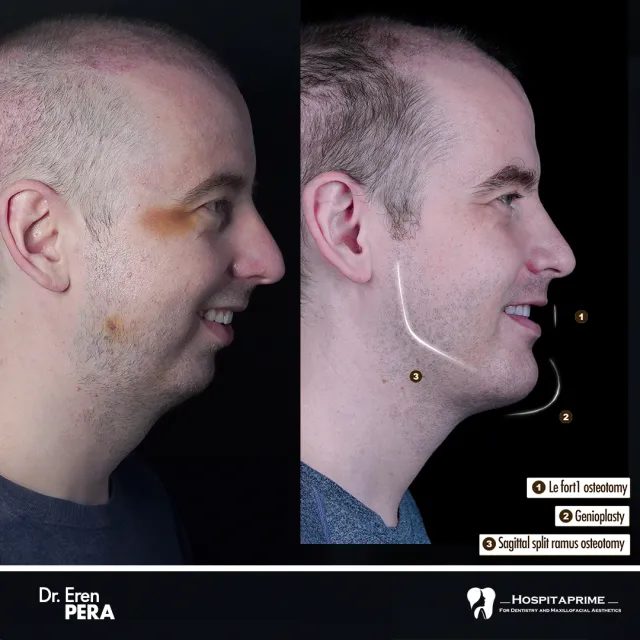
Most users on this forum understand the basics of a LeFort 1 bimax or the midface advancement of a LeFort 3. However, if you want to understand the absolute architectural limit of human skull modification, you must study the LeFort 4 osteotomy.
This is not a cosmetic procedure. It is an extreme, highly complex transcranial operation performed almost exclusively on pediatric and adult patients suffering from severe syndromic craniosynostosis (such as Crouzon, Apert, or Pfeiffer syndromes).
This guide serves as an anatomical, clinical, and financial deep-dive into the ultimate frontier of craniofacial engineering.
1. Structural Comparison: LeFort I, II, III vs. IV
Understanding where the cuts are made highlights why the LeFort 4 exists in its own separate risk tier.
2. The Surgical Mechanics: Step-by-Step Craniofacial Dissociation
A LeFort 4 requires a multidisciplinary surgical team, usually consisting of a pediatric neurosurgeon and a senior craniomaxillofacial surgeon.
3. Severe Clinical Risks and Complications
Because this surgery borders the anterior cranial fossa, the safety threshold is paper-thin. According to surgical reviews found on the Maastricht University Research Portal and StatPearls, complex midface osteotomies introduce dangerous physiological vulnerabilities:
4. Regional Pricing and Healthcare Realities
Because a LeFort 4 is classified as an intensive, reconstructive craniomaxillofacial surgery rather than a cosmetic procedure, it is rarely priced as an out-of-pocket, flat-fee operation. Most data is derived from institutional billing across different regions:
5. World-Class Surgeons and Major Treatment Hubs
No commercial "looksmaxing" surgeon handles this operation. The global authorities are highly academic research institutions:
6. The Blackpill Takeaway: Replicating the Aesthetics Safely
The LeFort 4 proves that your forehead slope, eye socket depth, and jaw position are legally locked together by a single macro-bone structure.
If you are a regular person looking to replicate these extreme structural shifts without risking death or blindness, you must use a modular approach combining multiple safe procedures:
7. Verifiable Medical Literature & References
To verify the biomechanics discussed in this thread, reference these official publications:
This is not a cosmetic procedure. It is an extreme, highly complex transcranial operation performed almost exclusively on pediatric and adult patients suffering from severe syndromic craniosynostosis (such as Crouzon, Apert, or Pfeiffer syndromes).
This guide serves as an anatomical, clinical, and financial deep-dive into the ultimate frontier of craniofacial engineering.
1. Structural Comparison: LeFort I, II, III vs. IV
Understanding where the cuts are made highlights why the LeFort 4 exists in its own separate risk tier.
| Procedure | Anatomical Cut Lines | Primary Aesthetic/Functional Impact |
|---|---|---|
| LeFort I | Separation of the maxilla from the pterygoid plates, just above the tooth roots. | Repositions the dental arch, fixes malocclusion, and alters subnasal/maxillary projection. |
| LeFort II | Pyramidal cut spanning across the nasal bridge down through the medial orbital floor. | Corrects central midface recession and severe paranasal deficiency. |
| LeFort III | Complete separation of the midface (zygomas, nose, orbit floor) from the cranium. | Eradicates massive midface hypoplasia, expands orbital volume, and fixes severe "bug eyes". |
| LeFort IV | Transcranial cut spanning the frontal bone (forehead), the nasofrontal suture, and the entire lateral and medial orbital walls. | Moves the forehead, brow ridge, eye sockets, nose, cheekbones, and upper jaw forward as a single massive unit. |
2. The Surgical Mechanics: Step-by-Step Craniofacial Dissociation
A LeFort 4 requires a multidisciplinary surgical team, usually consisting of a pediatric neurosurgeon and a senior craniomaxillofacial surgeon.
- Step 1: The Coronal Incision. The surgeon makes an incision across the scalp from ear to ear, peeling the entire face and forehead skin down to expose the bare skull.
- Step 2: The Frontal Craniotomy. The neurosurgeon carefully removes a piece of the frontal bone (forehead) to expose the frontal lobe of the brain. The brain is gently retracted to protect it during the deeper cuts.
- Step 3: The Subcranial Osteotomies. The surgeon cuts completely across the roof of the eye sockets (orbital roofs) and down through the lateral walls.
- Step 4: Pterygomaxillary Separation. The upper jaw is separated from the skull base at the back of the mouth. At this point, the entire facial skeleton and forehead are fully un-anchored from the braincase.
- Step 5: Distraction Osteogenesis (The Extension Phase). Rigid metal pins are anchored into the stable cranium, and internal or external distraction devices (like a halo frame) are attached to the detached face. Over several weeks, the hardware is turned 1mm per day to slowly pull the entire face forward, forcing new bone to grow into the widening gap.
3. Severe Clinical Risks and Complications
Because this surgery borders the anterior cranial fossa, the safety threshold is paper-thin. According to surgical reviews found on the Maastricht University Research Portal and StatPearls, complex midface osteotomies introduce dangerous physiological vulnerabilities:
- Cerebrospinal Fluid (CSF) Leaks: Dropping the cut across the cribriform plate can puncture the dura mater surrounding the brain, causing brain fluid to leak out of the nose.
- Meningitis and Encephalitis: Exposure of the brain to the nasal passages opens a direct path for aggressive bacterial infections.
- Total Visual Loss: Bone cuts passing millimeters away from the optic nerve carry a risk of permanent blindness.
- Severe Vascular Hemorrhage: Disruption of the internal maxillary artery or the pterygoid venous plexus can lead to rapid, life-threatening blood loss.
4. Regional Pricing and Healthcare Realities
Because a LeFort 4 is classified as an intensive, reconstructive craniomaxillofacial surgery rather than a cosmetic procedure, it is rarely priced as an out-of-pocket, flat-fee operation. Most data is derived from institutional billing across different regions:
- United States: $120,000 – $250,000+
- Context: This requires extended ICU stays, multiple custom 3D distraction devices, a neurosurgeon, and an orthopedic team. It is almost entirely paid for via medical insurance if a genetic syndrome is documented.
- Western Europe (UK, Germany, France): Fully funded by public healthcare systems (NHS / Social Security)
- Context: Provided exclusively via specialized state-run university hospitals for syndromic patients.
- South Korea & Taiwan: $60,000 – $90,000
- Context: Performed at major world-class medical hubs (like Chang Gung Memorial Hospital) for reconstructive cases.
5. World-Class Surgeons and Major Treatment Hubs
No commercial "looksmaxing" surgeon handles this operation. The global authorities are highly academic research institutions:
- Dr. Yoshiaki Sakamoto (Japan): Famous for pioneering internal distraction devices specifically optimized for LeFort IV + I combined procedures at elite university clinics.
- Seattle Children’s Craniofacial Center (USA): Home to world-class teams specializing in complex Counterclockwise Craniofacial Distraction Osteogenesis (C3DO) and total facial advancements.
- Texas Children’s Hospital / Craniofacial Team (USA): Internationally recognized for handling extensive LeFort advancements and custom segmentation techniques to correct severe skull dysmorphia.
6. The Blackpill Takeaway: Replicating the Aesthetics Safely
The LeFort 4 proves that your forehead slope, eye socket depth, and jaw position are legally locked together by a single macro-bone structure.
If you are a regular person looking to replicate these extreme structural shifts without risking death or blindness, you must use a modular approach combining multiple safe procedures:
- Custom PEEK/Titanium Forehead Implants: To manually build a flat or masculine brow ridge.
- Infraorbital-Malar Implants: Placed over the cheekbones to pull the lower eye socket structure forward and eliminate a recessed midface appearance.
- LeFort I Bimaxillary Advancement: To move your lower dental arch forward, providing the structural project your mouth needs without disturbing your brain cavity.
7. Verifiable Medical Literature & References
To verify the biomechanics discussed in this thread, reference these official publications:
- Distraction Mechanics: Look at Denny et al.'s foundational work on Rotation advancement of the midface by distraction osteogenesis via the National Center for Biotechnology Information (NCBI).
- Internal Fixation Guides: Read the comprehensive clinical breakdown on Le Fort Osteotomy Protocols provided by StatPearls.