

Stop Bonesmashing with Hammers: How to ACTUALLY Force Craniofacial Bone Deposition (Wolff's Law + Periost | [SWITCH TO DARK MODE]

khhvWarrior00

khhvWarrior00

self made
Joined
Aug 30, 2026
Posts
67
Reputation
46

Switch to dark mode for better experience


Disclaimer: This guide is strictly for educational, scientific, and biomechanical analysis. It covers skeletal histology, osteoblast and osteoclast signaling pathways, and Wolff Law mechanisms. I am not a physician and this does not constitute medical advice.


:what:Acknowledge the grey status before replying. This is a fresh account, I lost login for my main. Skip the insults and read the actual osteogenesis pathways below:

INTRODUCTION AND THE GREYCEL FALLACY

The aesthetics community is plagued by two equally low IQ extremes when it comes to bone structure:

  1. The TikTok Hammer Coper: Thinks hitting their zygomatic arches with blunt force (bonesmashing) causes controlled bone expansion, oblivious to the fact that it only causes subcutaneous hematomas, fat necrosis, and irregular bone resorption.
  2. The Blackpilled Nihilist: Claims that past age 18, bone tissue is 100 percent static and zero structural changes can occur without full osteotomies such as BSO, LeFort, or custom implants.
Both positions ignore basic skeletal biology. Craniofacial bones are dynamic, highly vascularized living structures governed by Mechanotransduction—the cellular process by which physical strain is converted into biochemical signals that drive Osteogenesis (bone deposition).

This guide covers the exact biochemical pathways, mechanical force vectors, and nutritional cofactors required to force localized bone remodeling along the mandible, maxilla, and orbital rims.

1. HISTOLOGY AND CELLULAR BIOLOGY OF BONE REMODELING

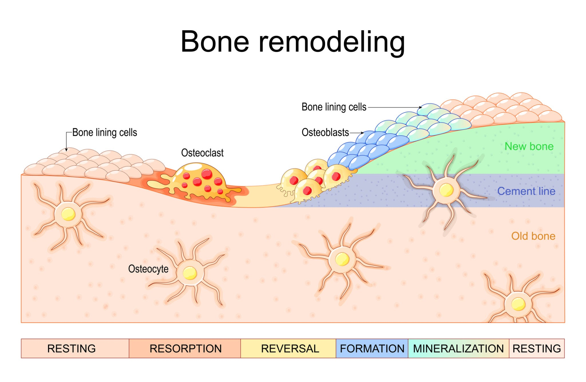
To force bone deposition, you must understand the Basic Multicellular Unit (BMU) operating within the Periosteum (the dense membrane covering the outer surface of bones).

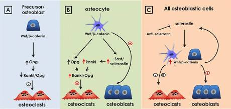
[MECHANICAL STRAIN OR TENSION]


[OSTEOCYTE FLUID SHEAR STRESS] ── Suppresses Sclerostin (SOST)


[RUNX2 and Wnt / Beta Catenin Activation]


[OSTEOBLAST PROLIFERATION] ── Secretes Type I Collagen Matrix (Osteoid)


[MINERALIZATION VIA OSTEOCALCIN] ── Solid Bone Deposition (Hydroxyapatite)


1788342030026

Key Biological Concepts:​

  • Osteocytes as Strain Sensors: Osteocytes reside inside microscopic cavities called lacunae within the bone matrix. When mechanical load flexes the bone, fluid moves through the lacunar canalicular network. Osteocytes sense this fluid shear stress via Piezo1 and Piezo2 mechanosensitive ion channels.
  • Sclerostin Suppression: Under zero strain, osteocytes release Sclerostin (encoded by the SOST gene), a protein that actively blocks bone formation. Applying mechanical strain suppresses Sclerostin, pulling the parking brake off bone growth.
  • The Wnt / Beta Catenin Pathway: Once Sclerostin is suppressed, the Wnt signaling pathway activates, forcing undifferentiated mesenchymal stem cells in the periosteum to transform into active Osteoblasts (bone building cells).
  • Osteoid Matrix Deposition: Osteoblasts synthesize an unmineralized organic matrix composed primarily of Type I Collagen (Osteoid). Over a period of 30 to 90 days, this matrix absorbs calcium and phosphate to crystallize into solid Hydroxyapatite.

2. INTRAMEMBRANOUS VS ENDOCHONDRAL VS PERIOSTEAL REMODELING

Understanding where and how different facial bones expand requires distinguishing between the three primary modes of skeletal growth:

Intramembranous Ossification​

Flat bones of the cranium, maxilla, and zygoma develop directly from mesenchymal connective tissue without a cartilage precursor. The periosteal membrane surrounding these bones retains lifelong osteogenic potential. When continuous tensile force is applied to periosteal fibers, osteoblasts deposit new bone layers directly onto the outer cortical shell (appositional growth).

Endochondral Ossification​

The mandibular condyles grow via a cartilage intermediate. During puberty, Growth Hormone Schemat mechanotransdukcji w kosciachnd IGF1 drive rapid chondrocyte proliferation at the condylar head, increasing total mandibular length and ramus height.

Craniofacial Suture Mechanics​

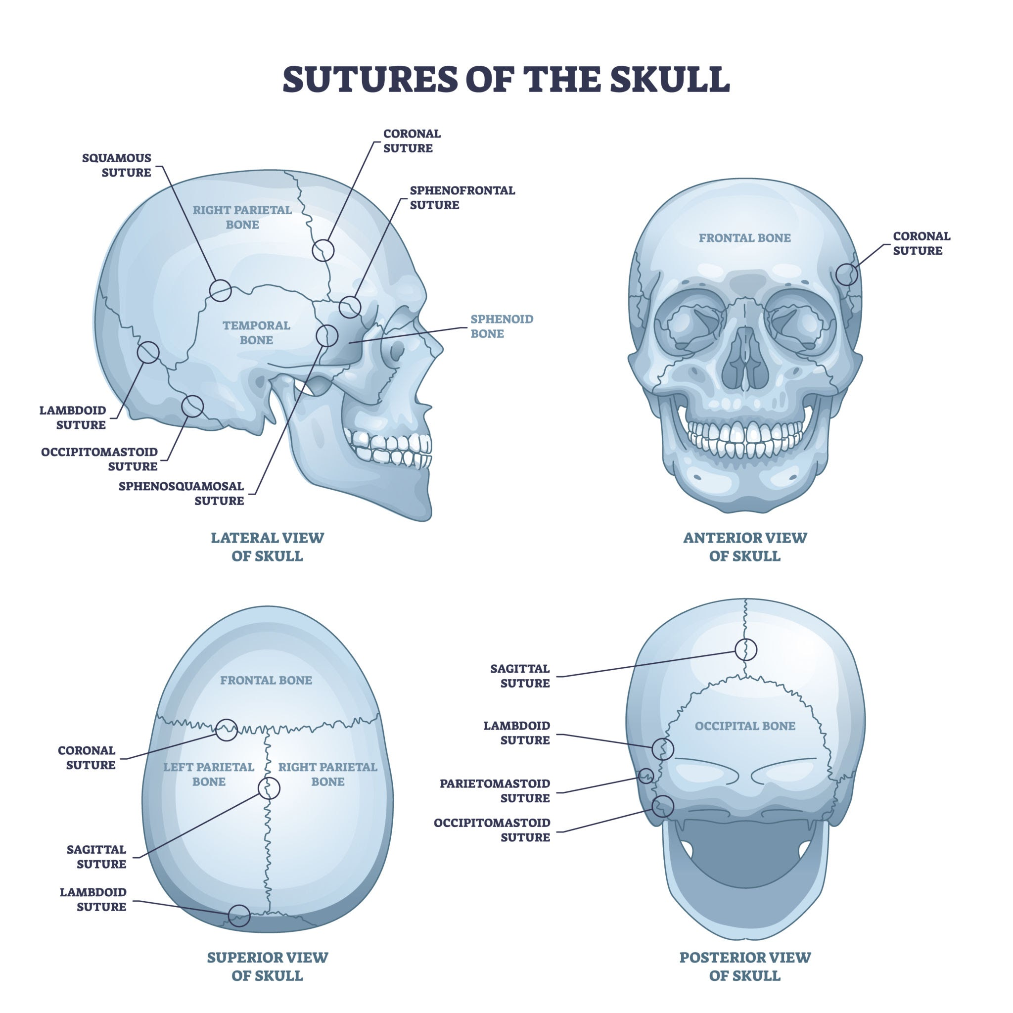
Facial sutures (such as the midpalatal suture, zygomaticomaxillary suture, and spheno occipital synchondrosis) are fibrous joints filled with osteogenic stem cells. While patent during prepubertal and pubertal development, applying outward mechanical tension across these sutures forces rapid osteoblast proliferation, permanently expanding facial width.
1788341845143

3. LOWER THIRD STRUCTURAL MECHANICS: MANDIBLE, RAMUS AND GONIAL ANGLE

The mandible is the most mechanically responsive bone in the craniofacial complex because it is attached to the strongest masticatory muscle: the Masseter.

Mechanosensation Protocol for Lower Third Widening:​

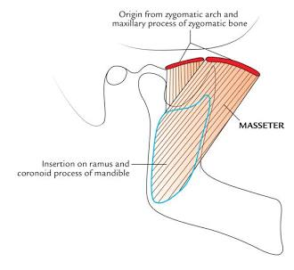
The masseter muscle inserts directly into the periosteum of the gonial angle and lateral ramus. Heavy muscular contractions apply direct mechanical traction and tension on the periosteum, signaling osteoblasts to deposit bone along the angle over time.
1788342059112
  1. Resistance Loading: Use dental grade mastic resin or dense gum.
  2. Duty Cycle: 30 to 45 minutes every 48 hours. Do not chew daily. Bone periosteum requires recovery periods for osteoid matrix secretion.
  3. Bite Vector: Keep your incisors aligned and bite down evenly on the molars and premolars to distribute equal mechanical shear stress across both mandibles, preventing asymmetry.
  4. Occlusion Protection: Ensure proper jaw alignment to prevent Temporomandibular Joint (TMJ) strain. Stop immediately if clicking or joint pain occurs.

4. MIDFACE AND UPPER THIRD STRUCTURAL MECHANICS: MAXILLA, ZYGOMA AND BROW RIDGE

The maxilla forms the central pillar of facial attractiveness, supporting the upper lip, nose, and infraorbital rims. While main midface sutures slow down their expansion velocity post puberty, surface periosteal remodeling remains active throughout life.

The Palatal Mechano Pressure Protocol:​

  • Posterior Palatal Load (Proper Mewing): The posterior third of the tongue must exert continuous upward and forward isometric force against the hard palate.
  • Fluid Shear Stress Mechanism: This continuous pressure creates mechanical strain across the palatine process and maxilla. Osteocytes in the infraorbital rim detect this pressure, signaling osteoblasts to prevent midface bone resorption and maintain maxilla support under the eyes.

Zygomatic Arch Expansion:​

The zygomaticus major and masseter muscles pull directly on the zygomatic arch. High load mechanical chewing combined with isometric facial tension increases periosteal shear stress along the cheekbone arch, encouraging subtle appositional thickening over extended multi month cycles.

5. THE ENDOCRINE AND PHARMACOLOGICAL MATRIX FOR OSTEOGENESIS

Creating mechanical strain without saturating your systemic hormonal pool will yield minimal structural changes. You must optimize the endocrine environment for bone matrix deposition.

1. Growth Hormone (GH) and Hepatic IGF1​

GH stimulates pre chondrocytes, while IGF1 (Insulin like Growth Factor 1) directly drives osteoblast proliferation and collagen matrix synthesis. Elevate natural GH pulses via deep slow wave sleep, cold exposure, and high intensity compound weightlifting.

2. Dihydrotestosterone (DHT) vs Testosterone​

Testosterone increases general bone density, but DHT exhibits a significantly higher binding affinity for androgen receptors in facial periosteal tissue. DHT promotes masculine bone density, thickening the supraorbital ridge, mandible, and chin without aromatizing into estrogen.

3. Estrogen Control and Aromatase Inhibition​

While baseline estrogen is required for bone mineral density, elevated Estradiol accelerates epiphyseal plate fusion and sutural closure. Keeping body fat between 10 to 14 percent minimizes aromatase density, preserving open growth windows and delaying sutural calcification.

4. Thyroid Axis​

Active Triiodothyronine regulates the metabolic rate of bone turnover. Subclinical hypothyroidism slows down osteoblast matrix deposition, resulting in delayed bone mineralization.

6. THE OSTEO MINERAL AND COFACTOR STACK

If you stimulate osteoblasts without providing raw mineral substrates, your body cannot mineralize the soft osteoid matrix into hard hydroxyapatite.
1788342114475

7. MASTER EXECUTION BLUEPRINT AND TIMELINE

Bone remodeling is a slow, structural process governed by cellular turnover rates. Attempting to rush this via extreme blunt force will fail.

MONTHS 1 THROUGH 2: Endocrine Preparation and Mineral Saturation
* Establish nutrient stack (Vit D3, K2, MCHA, Boron, Magnesium).
* Drop body fat to 10 to 14 percent to suppress aromatase.

MONTHS 3 THROUGH 6: Mechanical Load and Periosteal Strain Activation
* Initiate mastic resin chewing protocol (30 to 45 mins every second day).
* Maintain continuous posterior palatal tongue pressure.

MONTHS 7 THROUGH 12: Osteoid Calcification and Solidification
* Osteoblasts deposit Type I collagen matrix along strained periosteal zones.
* Hydroxyapatite crystals solidify new cortical bone layers.

8. FREQUENTLY ASKED QUESTIONS AND FAILURE MODES

Why Bonesmashing is Low IQ:

Hitting the face with hard objects creates uncontrolled microfractures, subcutaneous hematomas, and soft tissue scarring. When bone suffers severe acute trauma without controlled directional strain, osteoclasts flood the area to clear damaged tissue, often leading to irregular bone resorption (making the bone smaller or uneven) rather than smooth appositional growth.

Correcting Facial Asymmetry:

If one side of your mandible or maxilla is less developed, check your mastication habits and sleeping posture. Always load the weaker side with equal or slightly higher mechanical duty cycles while maintaining proper spinal and neck alignment.

Preventing TMJ Injury:

Never chew through sharp joint pain. The temporomandibular joint contains a delicate fibrocartilaginous disc. If you experience clicking or popping, immediately reduce chewing resistance, focus on soft tissue massage of the pterygoid muscles, and resume only when fully asymptomatic.


It took me 3 hours:soy::smonk:
 
  • +1
Reactions: Mazilias, Absurdist and 76.1
Hammers my cock to shit n piss tbh
 
  • +1
  • JFL
Reactions: khhvWarrior00, Mazilias and 76.1
read every single molecule
 
  • +1
Reactions: khhvWarrior00, Absurdist and 76.1
Will read later. Nice effort.
 
  • JFL
Reactions: ZyzzReincarnate
Switch to dark mode for better experience


Disclaimer: This guide is strictly for educational, scientific, and biomechanical analysis. It covers skeletal histology, osteoblast and osteoclast signaling pathways, and Wolff Law mechanisms. I am not a physician and this does not constitute medical advice.


:what:Acknowledge the grey status before replying. This is a fresh account, I lost login for my main. Skip the insults and read the actual osteogenesis pathways below:

INTRODUCTION AND THE GREYCEL FALLACY

The aesthetics community is plagued by two equally low IQ extremes when it comes to bone structure:

  1. The TikTok Hammer Coper: Thinks hitting their zygomatic arches with blunt force (bonesmashing) causes controlled bone expansion, oblivious to the fact that it only causes subcutaneous hematomas, fat necrosis, and irregular bone resorption.
  2. The Blackpilled Nihilist: Claims that past age 18, bone tissue is 100 percent static and zero structural changes can occur without full osteotomies such as BSO, LeFort, or custom implants.
Both positions ignore basic skeletal biology. Craniofacial bones are dynamic, highly vascularized living structures governed by Mechanotransduction—the cellular process by which physical strain is converted into biochemical signals that drive Osteogenesis (bone deposition).

This guide covers the exact biochemical pathways, mechanical force vectors, and nutritional cofactors required to force localized bone remodeling along the mandible, maxilla, and orbital rims.

1. HISTOLOGY AND CELLULAR BIOLOGY OF BONE REMODELING

To force bone deposition, you must understand the Basic Multicellular Unit (BMU) operating within the Periosteum (the dense membrane covering the outer surface of bones).

[MECHANICAL STRAIN OR TENSION]


[OSTEOCYTE FLUID SHEAR STRESS] ── Suppresses Sclerostin (SOST)


[RUNX2 and Wnt / Beta Catenin Activation]


[OSTEOBLAST PROLIFERATION] ── Secretes Type I Collagen Matrix (Osteoid)


[MINERALIZATION VIA OSTEOCALCIN] ── Solid Bone Deposition (Hydroxyapatite)


View attachment 5596829

Key Biological Concepts:​

  • Osteocytes as Strain Sensors: Osteocytes reside inside microscopic cavities called lacunae within the bone matrix. When mechanical load flexes the bone, fluid moves through the lacunar canalicular network. Osteocytes sense this fluid shear stress via Piezo1 and Piezo2 mechanosensitive ion channels.
  • Sclerostin Suppression: Under zero strain, osteocytes release Sclerostin (encoded by the SOST gene), a protein that actively blocks bone formation. Applying mechanical strain suppresses Sclerostin, pulling the parking brake off bone growth.
  • The Wnt / Beta Catenin Pathway: Once Sclerostin is suppressed, the Wnt signaling pathway activates, forcing undifferentiated mesenchymal stem cells in the periosteum to transform into active Osteoblasts (bone building cells).
  • Osteoid Matrix Deposition: Osteoblasts synthesize an unmineralized organic matrix composed primarily of Type I Collagen (Osteoid). Over a period of 30 to 90 days, this matrix absorbs calcium and phosphate to crystallize into solid Hydroxyapatite.

2. INTRAMEMBRANOUS VS ENDOCHONDRAL VS PERIOSTEAL REMODELING

Understanding where and how different facial bones expand requires distinguishing between the three primary modes of skeletal growth:

Intramembranous Ossification​

Flat bones of the cranium, maxilla, and zygoma develop directly from mesenchymal connective tissue without a cartilage precursor. The periosteal membrane surrounding these bones retains lifelong osteogenic potential. When continuous tensile force is applied to periosteal fibers, osteoblasts deposit new bone layers directly onto the outer cortical shell (appositional growth).

Endochondral Ossification​

The mandibular condyles grow via a cartilage intermediate. During puberty, Growth Hormone Schemat mechanotransdukcji w kosciachnd IGF1 drive rapid chondrocyte proliferation at the condylar head, increasing total mandibular length and ramus height.

Craniofacial Suture Mechanics​

Facial sutures (such as the midpalatal suture, zygomaticomaxillary suture, and spheno occipital synchondrosis) are fibrous joints filled with osteogenic stem cells. While patent during prepubertal and pubertal development, applying outward mechanical tension across these sutures forces rapid osteoblast proliferation, permanently expanding facial width.

3. LOWER THIRD STRUCTURAL MECHANICS: MANDIBLE, RAMUS AND GONIAL ANGLE

The mandible is the most mechanically responsive bone in the craniofacial complex because it is attached to the strongest masticatory muscle: the Masseter.

Mechanosensation Protocol for Lower Third Widening:​

The masseter muscle inserts directly into the periosteum of the gonial angle and lateral ramus. Heavy muscular contractions apply direct mechanical traction and tension on the periosteum, signaling osteoblasts to deposit bone along the angle over time.
  1. Resistance Loading: Use dental grade mastic resin or dense gum.
  2. Duty Cycle: 30 to 45 minutes every 48 hours. Do not chew daily. Bone periosteum requires recovery periods for osteoid matrix secretion.
  3. Bite Vector: Keep your incisors aligned and bite down evenly on the molars and premolars to distribute equal mechanical shear stress across both mandibles, preventing asymmetry.
  4. Occlusion Protection: Ensure proper jaw alignment to prevent Temporomandibular Joint (TMJ) strain. Stop immediately if clicking or joint pain occurs.

4. MIDFACE AND UPPER THIRD STRUCTURAL MECHANICS: MAXILLA, ZYGOMA AND BROW RIDGE

The maxilla forms the central pillar of facial attractiveness, supporting the upper lip, nose, and infraorbital rims. While main midface sutures slow down their expansion velocity post puberty, surface periosteal remodeling remains active throughout life.

The Palatal Mechano Pressure Protocol:​

  • Posterior Palatal Load (Proper Mewing): The posterior third of the tongue must exert continuous upward and forward isometric force against the hard palate.
  • Fluid Shear Stress Mechanism: This continuous pressure creates mechanical strain across the palatine process and maxilla. Osteocytes in the infraorbital rim detect this pressure, signaling osteoblasts to prevent midface bone resorption and maintain maxilla support under the eyes.

Zygomatic Arch Expansion:​

The zygomaticus major and masseter muscles pull directly on the zygomatic arch. High load mechanical chewing combined with isometric facial tension increases periosteal shear stress along the cheekbone arch, encouraging subtle appositional thickening over extended multi month cycles.

5. THE ENDOCRINE AND PHARMACOLOGICAL MATRIX FOR OSTEOGENESIS

Creating mechanical strain without saturating your systemic hormonal pool will yield minimal structural changes. You must optimize the endocrine environment for bone matrix deposition.

1. Growth Hormone (GH) and Hepatic IGF1​

GH stimulates pre chondrocytes, while IGF1 (Insulin like Growth Factor 1) directly drives osteoblast proliferation and collagen matrix synthesis. Elevate natural GH pulses via deep slow wave sleep, cold exposure, and high intensity compound weightlifting.

2. Dihydrotestosterone (DHT) vs Testosterone​

Testosterone increases general bone density, but DHT exhibits a significantly higher binding affinity for androgen receptors in facial periosteal tissue. DHT promotes masculine bone density, thickening the supraorbital ridge, mandible, and chin without aromatizing into estrogen.

3. Estrogen Control and Aromatase Inhibition​

While baseline estrogen is required for bone mineral density, elevated Estradiol accelerates epiphyseal plate fusion and sutural closure. Keeping body fat between 10 to 14 percent minimizes aromatase density, preserving open growth windows and delaying sutural calcification.

4. Thyroid Axis​

Active Triiodothyronine regulates the metabolic rate of bone turnover. Subclinical hypothyroidism slows down osteoblast matrix deposition, resulting in delayed bone mineralization.

6. THE OSTEO MINERAL AND COFACTOR STACK

If you stimulate osteoblasts without providing raw mineral substrates, your body cannot mineralize the soft osteoid matrix into hard hydroxyapatite.

7. MASTER EXECUTION BLUEPRINT AND TIMELINE

Bone remodeling is a slow, structural process governed by cellular turnover rates. Attempting to rush this via extreme blunt force will fail.

MONTHS 1 THROUGH 2: Endocrine Preparation and Mineral Saturation
* Establish nutrient stack (Vit D3, K2, MCHA, Boron, Magnesium).
* Drop body fat to 10 to 14 percent to suppress aromatase.

MONTHS 3 THROUGH 6: Mechanical Load and Periosteal Strain Activation
* Initiate mastic resin chewing protocol (30 to 45 mins every second day).
* Maintain continuous posterior palatal tongue pressure.

MONTHS 7 THROUGH 12: Osteoid Calcification and Solidification
* Osteoblasts deposit Type I collagen matrix along strained periosteal zones.
* Hydroxyapatite crystals solidify new cortical bone layers.

8. FREQUENTLY ASKED QUESTIONS AND FAILURE MODES

Why Bonesmashing is Low IQ:

Hitting the face with hard objects creates uncontrolled microfractures, subcutaneous hematomas, and soft tissue scarring. When bone suffers severe acute trauma without controlled directional strain, osteoclasts flood the area to clear damaged tissue, often leading to irregular bone resorption (making the bone smaller or uneven) rather than smooth appositional growth.

Correcting Facial Asymmetry:

If one side of your mandible or maxilla is less developed, check your mastication habits and sleeping posture. Always load the weaker side with equal or slightly higher mechanical duty cycles while maintaining proper spinal and neck alignment.

Preventing TMJ Injury:

Never chew through sharp joint pain. The temporomandibular joint contains a delicate fibrocartilaginous disc. If you experience clicking or popping, immediately reduce chewing resistance, focus on soft tissue massage of the pterygoid muscles, and resume only when fully asymptomatic.


It took me 3 hours:soy::smonk:
Just bonesmash man
 

Similar threads

uunltd
Replies
0
Views
32
uunltd
uunltd
Eldian
Replies
18
Views
161
zennn
zennn
matviww
Replies
10
Views
106
UnGhosted
UnGhosted
E
Replies
6
Views
70
Lowtiertroll
Lowtiertroll

Users who are viewing this thread

Back
Top