

tension
Luminary
- Joined
- Jan 18, 2026
- Posts
- 5,781
- Reputation
- 8,793
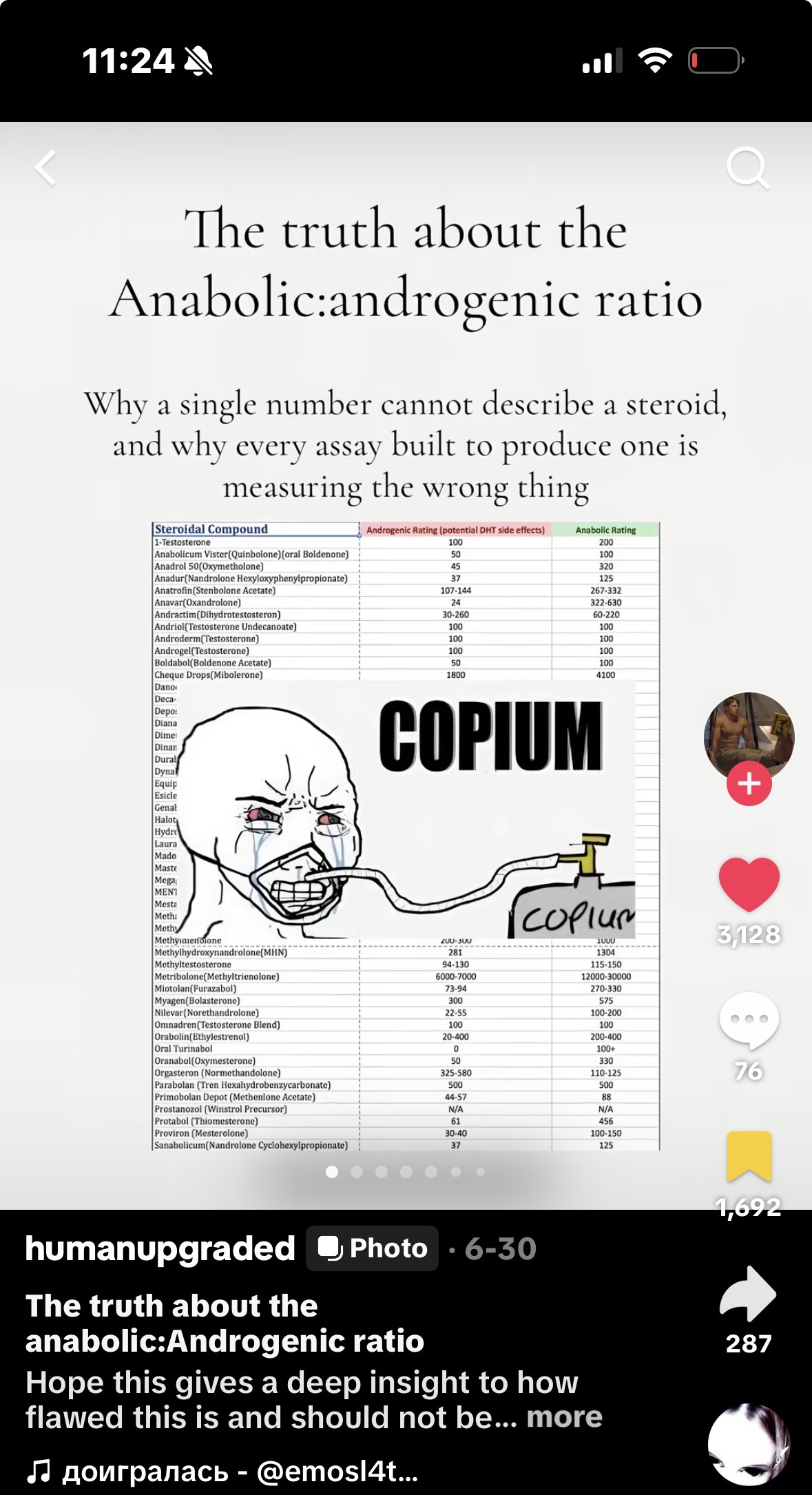
Why a single number cannot describe a steroid, and why every assay built to produce one is measuring the wrong thing
The anabolic-to-androgenic ratio is simply an attempt to express various compounds' degree of effectiveness on both the anabolic and androgenic axes. As such, we select a reference compound that will serve as our baseline - typically testosterone with an anabolic-to-androgenic ratio of 100:100 (unity), and compare each compound's anabolic-to-androgenic ratio to that of our selected reference compound. A steroid reported as 400:200 is, on this scheme, four times as anabolic and twice as androgenic as testosterone, molecule for molecule. In so doing, we are comparing, molecule for molecule, how effective each compound is when compared to testosterone.
The attraction is obvious. In other words, if there is a "clean" number separating the desired from undesired, then minimising the undesirable side effects will boil down to selecting a compound which has an optimal ratio. However, the number is bad prior to when any animals are given the drug or any receptors are titrated. The idea of the concept fails at the point of definition and the experimental methods developed upon this broken idea have their own inherent faults.
One of the first problems with the conceptualisation of androgenic is that it is not a single axis. It is a basket of different effects across different tissues that do not all move together. The most clear example of this is the prostate, which has a response to androgen that saturates within the low-hypogonadal range
“Prostate cancer growth is exquisitely sensitive to variations in androgen concentrations at or below the near-castrate range and is insensitive to variations above this concentration… Further increases in androgen concentration do not result in any further binding to AR.”
- Morgentaler & Traish (2009), European Urology 55:310–321
Half-maximal prostate development happens to occur approximately at 36 ng/dL (or ~1 nM) above which the androgen receptor is saturated [1] consistent with the fact that prostate volume is unaffected in normal males when they receive a dose of 600mg of testosterone enanthate each week for 20 weeks [7]; however, at this dose, there is an evident rise in all other androgenic effects including hematocrit, axis suppression, and sebaceous activity. It would be contradictory to fold multiple tissues into a singular "androgenic" axis; some of these tissues will plateau first while others will continue to increase proportionally as dosage increases.
The second problem is still deeper. Even granting a clean androgenic axis, scalar values cannot capture what they are supposed to summarise. Response of tissue to steroids is a surface in at least three dimensions: it depends on tissue type, concentration levels and time. Different tissues occupy different parts of this surface at different doses. Reducing a surface to a single point is a category error, independent of quality of measurement. The assays when examined below do not fail just because they are imprecise; they fail because the quantity they estimate does not exist in the form ratio takes.
The levator ani muscle is used as the index of anabolic effect, and the ventral prostate and seminal vesicles are used as the index of androgenic effect. The ratio of the increase in weight of the levator ani muscle compared to the increase in weight of the accessory sex organs determines the anabolic/androgenic ratio.
While even based upon these internal criteria alone, the method has an inherent instability due to different dose response curves being present for both muscle and sex organs. This was noted by Hershberger himself:
“The slope of the levator ani dose-response curve does not parallel those for the accessory glands; the curves for ventral prostate and seminal vesicles are approximately parallel.”
- Hershberger, Shipley & Meyer (1953), Proc. Soc. Exp. Biol. Med. 83:175–180
Non-parallel curves mean that the ratio of the two readouts is not a constant for all dose levels within the compound. You report your number wherever on the two curves you happen to be taking data from.
The critical flaw is with the numerator. It's worth exploring this further because it's often glossed over. The "muscle" treated by the assay as a surrogate for skeletal muscle isn't actually a surrogate; it's a misnomer.
“Although the rat’s ‘levator ani’ muscle has been widely used for screening and evaluating nonandrogenic anabolic steroids… this muscle is not homologous to the one called levator ani in man, cat, and dog, but is actually the dorsal bulbocavernosus, a muscle peculiar to rodents and… clearly part of the male reproductive system.” - Hayes (1965), Acta Endocrinologica 48:337–347
This is a perineal muscle of the male genital apparatus which encircles the root of the penis and is not a posture or locomotion muscle nor does it have a functionally related relationship to the quadriceps, pectoralis or any other muscle a clinician would treat for wasting or an athlete attempting to develop would find useful. The name has been a two century-long anatomical mistake that has been passed down by this assay.
Its existence is driven by androgens. The bulbocavernosus muscle and its motor neurons represent a sexually dimorphic neuromuscular system that includes the spinal nucleus of the bulbocavernosus (SNB) which requires perinatal androgen exposure to survive during development [5]. Females and androgen-insensitive (Tfm) rats/animals generally do not possess the muscle or motor pool of this muscle. Its entire developmental program is dictated by androgens, a characteristic that no limb muscle possesses. Treating growth of the muscle as a general indicator of "anabolism" is to mistakenly identify an accessory sex organ as skeletal muscularity.
It is far more androgen-responsive than locomotor muscle, and wastes like a denervated muscle when androgen is withdrawn. Rat limb/trunk muscle is relatively devoid of androgen receptors and obtains its mass primarily through mechanical load, satellite cell activity and the IGF-1/mTOR pathway. Perineal muscles behave similarly to their anatomically associated accessory sex organs. An electron microscopic study documenting the castrate response demonstrated this clearly: the dorsal bulbocavernosus falls to approximately 55% of control weight at 15 d, 39.5% at one mo., and 15.6% at eight mo. and:
“The rate of weight decrease of dorsal bulbocavernosus muscle after castration is similar to that of denervation atrophy in rat skeletal muscles.”
- Gori, Pellegrino & Pollera (1967), Experimental and Molecular Pathology 6:172–198
Real skeletal muscle does not collapse like a denervated muscle when androgen is lost. A muscle that exists solely based upon whether or not androgen is present, by design, reports androgen responsiveness not anabolism. (Hayes' group, similar to Hayes, abandoned the old name, stating in the abstract: “It should be correctly named dorsal bulbocavernosus. We will use this name throughout this write-up/slideshow.”)
Since there is no relevant human homologue, even a perfect measurement using rat results could not be transferred. The human muscles whose growth constitute the actual therapeutic and athletic targets do not exist in rat genital muscles used in this study. Hayes' summation regarding the whole methodology is worthy of citation in full:
“The so-called ‘levator ani’ of the rat has been misnamed, inaccurately dissected, incorrectly described, and misleadingly used. Its response to steroids is a masculinizing effect, and is accompanied by masculinization of other organs. Its response is not a reflection of general myotrophic action, and has not been shown to correlate highly with any clearly anabolic response… This investigation confirms the view that the ‘levator ani test’ is useless for either preliminary screening or quantitative evaluation of nonandrogenic anabolic steroids.”
- Hayes (1965), Acta Endocrinologica 48:337–347
When you combine these two parts, the way this test is structured with the errors becomes apparent. The "anabolic" portion of the results from the assay is also an androgenic tissue. It uses an androgenic response in the numerator and another androgenic response in the denominator, then shows the result of dividing them by one another as if it is a measure of anabolic activity over androgenic. When muscle was selected, the numerator was poisoned.
The rest of the problems stated above are true; however, they are secondary issues. The ratio is dependent on the dose and time. Because the seminal vesicles and bulbocavernosus will lose their growth differently depending on the concentration of the compounds, van der Vies demonstrated that for nandrolone, both tissues will increase while drug levels are elevated; however, when the depot is depleted, the seminal vesicles will have returned to baseline while the muscle will continue growing. His conclusion to his study states this issue perfectly:
“The dissociation of anabolic from androgenic effects depends on the moment of observation after administration of the compound. Secondly, different target organs respond in a different fashion to changes in the concentration of the stimulating agent in the course of time.”
- van der Vies (1985), Acta Endocrinologica Suppl. 271:38–43
He then isolated the variable with a simulation. He found that giving the same total dose of naked nandrolone as a single dose vs. spread over twenty-one doses resulted in differences in the ratio of levator ani to seminal vesicle weights, i.e., 3mg/2mg for a single dose, 34mg/34mg for an equivalent split-dose, and 30mg/17mg for nandrolone phenylpropionate [6]. Same molecule, same total dose; the ratio changed merely with varying dosing schedules. Additionally, there was also the assumption made without evidence that the growth of ventral prostate and seminal vesicles represented all aspects of androgenic action which the saturation behavior of the prostate already denies [1][7]; and finally, there was also the assumption made without testing that homologous human tissues react identically..
It would be reassuring to consider this simply a relic of history. It is not. The classic assay continues to be used today to validate modern selective androgen receptor modulators prior to entering clinical trials:
“The tissue selectivity of GSK2881078 was assessed in a classic Hershberger assay… [it] restored the weight of the levator ani muscle in orchiectomized rats to that of sham-operated rats but produced only a minor increase in prostate weight.”
- Neil et al. (2018), J. Clin. Endocrinol. Metab. 103:3215–3224
A 2018 development article, therefore, bases its "anabolic" claims entirely on the weight of a mis-named rat genital muscle.
Table 1. Relative binding affinities of selected steroids, after Saartok, Dahlberg & Gustafsson (1984); methyltrienolone is the reference (RBA = 1) for the muscle and prostate columns.
The most significant result in this report is not a number contained in Table 1 - it is a negative conclusion eliminating the rationale for an "anabolic" assay:
“In general the ligand specificity of the AR was the same in muscle and prostate. Hence, the existence of a specific anabolic receptor distinct from the AR must be seriously questioned.”
- Saartok, Dahlberg & Gustafsson (1984), Endocrinology 114:2100–2106
If muscle and prostate express the same receptor with identical ligand specificity, then "anabolic selectivity" cannot occur at the receptor. Any tissue selectivity exhibited by a steroid must arise from some process occurring after it binds to its target receptor (i.e., in tissue specific metabolism); however, this is beyond what can be observed in a binding assay. There are three specific examples demonstrating how these assays fail.
The results vary among species. Saartok's data demonstrate that 1α-methyl-DHT binds approximately three times weaker than testosterone in rat muscle; whereas 1α-methyl-DHT binds approximately three times stronger than testosterone in rabbit muscle. The direction of this result varies depending upon the species used to prepare the cytosol; since rat and rabbit cannot come into agreement regarding ranking order, there is no scientific basis for extrapolating either result to man.
Tissue metabolism is poorly characterised or completely absent - Saartok states this directly:
“Skeletal muscle differs from prostate in being virtually incapable of carrying out 5α-reduction… muscle has a T/DHT ratio about an order of magnitude higher than that of prostate… since DHT is rapidly inactivated in muscle, T is conceivably a more important receptor ligand than DHT in muscle.”
- Saartok, Dahlberg & Gustafsson (1984), Endocrinology 114:2100–2106
It is therefore the nature of the tissue, and not the receptor, that results in the low RBA of DHT in muscle. Muscle 3α-hydroxysteroid dehydrogenase reduces DHT to the non-ligand 3α-androstanediol [11], while Bergink et al. have shown the mirror image situation regarding nandrolone, as follows:
“The difference in anabolic and androgenic activity between nandrolone and testosterone is caused by the fact that 5α-reduction increases the affinity of T and decreases the affinity of nandrolone.”
- Bergink, Janssen, Turpijn & van der Vies (1985), J. Steroid Biochem. 22:831–836
Muscle tissue is able to convert testosterone into the more potent DHT via high levels of 5α-reductase. However, the same type of tissue can reduce nandrolone into the less potent dihydronandrolone via the action of the same 5α-reductase. Binding assays performed in either cytosols that do or do not perform such conversions will produce different ranking orders of the two compounds, while those performed using a cell-free system that performs neither conversion will produce incorrect ranking orders for both.
This is the identical limitation that all subsequent androgen responsive reporter (CALUX) bioassays inherited. CALUX bioassays represent a true advance beyond binding, as they measure transactivation rather than simply occupancy. Human osteosarcoma (U2-OS) cells were co-transfected with human androgen receptors (AR) and a firefly luciferase reporter gene driven by an androgen response element. Cells were then treated with a range of concentrations of the test compound and the resulting luminescence was used to fit a dose-response curve. From this curve, the relative potency (REP), expressed as the ratio of EC50 values for the reference compound divided by EC50 values for the test compound, was determined. As Houtman has indicated,
“The amount of luciferase produced is directly proportional to the amount and androgenic potency of the compound(s) the cells have been exposed to.”
- Houtman et al. (2009), Analytica Chimica Acta 637:247–258
However, this is receptor activation in a single transfected U2-OS cell line devoid of its endogenous steroid metabolising enzymes - precisely the biochemical steps involved in generating selectivity according to Saartok's argument. Therefore, the resultant values represent potency in a metabolic void.
Table 2. Relative potencies in receptor activation (REP) from the CALUX bioassay, after Houtman et al. (2009). DHT is the AR reference, ORG-2058 the PR reference, estradiol the ERα/β reference, and dexamethasone the GR reference. Values are normalised within each column, so they rank compounds within a receptor but cannot be compared across receptors.
In addition, binding affinity is also affected by plasma transport of steroids which is entirely missed by in vitro measurements. Saartok studied binding affinities of several steroids to human sex hormone-binding globulin (SHBG) in conjunction with their binding affinities to the AR and observed that the two types of affinities do not necessarily correlate e.g., 1α-Me-DHT binds approximately 4-fold more tightly to SHBG than DHT. They concluded that steroids could be grouped into separate categories based upon whether or not they bind to the AR, SHBG, both or neither, and that "the activities of these steroids in vivo would thus depend on both the affinity of the steroids for SHBG and the concentration of SHBG." Thus, a compound's free, biologically-active fraction in vivo depends upon a plasma protein that is not present in an assay dish.
Additionally, the technique violates the one it was intended to confirm and contradicts itself within individual studies. If one interprets the binding data correctly, testosterone appears to possess a superior anabolic-to-androgenic profile compared to nandrolone, i.e., the inverse of that demonstrated by Hershberger assay data. More seriously, Saartok reported that the in vivo ratios exhibited extreme variability for one compound:
“The in vivo anabolic-androgenic ratio varies in different studies from 0.4 to 1.9 for DHT, from 1.5 to 15 for 19-norT, from 1.8 to 10.6 for oxymetholone, and from 1.4 to 10.6 for stanozolol.”
- Saartok, Dahlberg & Gustafsson (1984), Endocrinology 114:2100–2106
If a given compound exhibits a ten-fold variation in its in vivo ratio across multiple studies, then it is clear that whatever quantity being measured is not related to properties of the compound. When there are two methods designed to measure the same quantity (i.e., two reference methods) and they yield conflicting conclusions (i.e., inverted conclusions) and one method demonstrates an order-of-magnitude variability in measurement for that quantity for the same compound across studies, then it is conclusive evidence that neither method is measuring some transferable invariant.
Therefore, the objective is not a ratio but rather a vector, specifically, a per-tissue profile of activity, and direct evidence supporting this mechanism exists in the clinical literature. In the 5α-Reductase Trial, suppressing testosterone's conversion to DHT by administering dutasteride failed to dampen gains in muscle mass or strength:
“Conversion of testosterone to DHT is not essential for mediating its anabolic effects on muscle… [DHT] amplifies the effects of testosterone in tissues with high 5α-reductase activity such as the prostate and skin, but not in tissues with low 5α-reductase activity such as skeletal muscle and bone.” - Bhasin et al. (2012), JAMA 307:931–939
It is crucial to note that this result included a qualification that must be explicitly mentioned and acknowledged: at supraphysiological testosterone concentrations employed during this study, testosterone alone was capable of maintaining prostate volume even though DHT was suppressed, thus, DHT amplification makes greatest sense when testosterone levels are low enough so that testosterone saturates prostate responsiveness. The amplification model and the saturation model are merely viewed from opposing vantage points.
Therefore, this study provides compelling evidence against selecting for potency solely based on binding affinity and argues for incorporating additional measures that take into account factors influencing activity after formation of steroid-protein complexes.
The fact that the muscle/prostate distinction has a metabolic cause that occurs in humans was not an inferential leap. Prostate tissue was cultured by Prout and his colleagues with labeled testosterone, and then they measured how much testosterone was converted into DHT. They found that, on average, BPH had converted about 61.7% of its testosterone to DHT, while human rectus muscle, actual human skeletal muscle, converted only about 8.3% of its testosterone to DHT. Even prostatic stroma converted significantly more testosterone to DHT than did human rectus muscle. Thus, human skeletal muscle is clearly a low-5α-reductase tissue; the prostate, skin, and scalp are all high-5α-reductase tissues [11]. That this metabolic geography is the total explanation for selective sensitivity to androgens as well.
Trenbolone makes the argument concrete. It has a 3-oxo-triene system which is poorly metabolised by 5 alpha reductase and therefore will not be converted to a more potent androgen in skin, scalp or prostate as testosterone is converted to DHT. Its genuine selectivity is created at one local enzymatic step – precisely the step that each of the Hershberger assay (incorrect tissue incorrect species), binding assays (no metabolism species dependent), and CALUX/REP bioassays (no metabolism one cell line) erased. These familiar figures quoting Trenbolone at somewhere near 500:500 are not just incorrect in magnitude – they were generated from systems structurally unable to see where Trenbolone’s selectivity actually exists. This is the lesson of the ratio in miniature: the number is not just incorrect in magnitude but was measured in the wrong location
2. Hershberger LG, Shipley EG, Meyer RK. Myotrophic activity of 19-nortestosterone and other steroids determined by modified levator ani muscle method. Proc Soc Exp Biol Med. 1953;83(1):175–180.
3. Gori Z, Pellegrino C, Pollera M. The castration atrophy of the dorsal bulbocavernosus muscle of rat: an electron microscopic study. Exp Mol Pathol. 1967;6(2):172–198.
4. Hayes KJ. The so-called ‘levator ani’ of the rat. Acta Endocrinol (Copenh). 1965;48(3):337–347.
5. Breedlove SM, Arnold AP. Hormone accumulation in a sexually dimorphic motor nucleus of the rat spinal cord. Science. 1980;210(4469):564–566.
6. van der Vies J. Implications of basic pharmacology in the therapy with esters of nandrolone. Acta Endocrinol (Copenh) Suppl. 1985;271:38–43.
7. Bhasin S, Travison TG, Storer TW, et al. Effect of testosterone supplementation with and without a dual 5α-reductase inhibitor on fat-free mass in men with suppressed testosterone production: a randomized controlled trial. JAMA. 2012;307(9):931–939.
8. Neil D, Clark RV, Magee M, et al. GSK2881078, a SARM, produces dose-dependent increases in lean mass in healthy older men and women. J Clin Endocrinol Metab. 2018;103(9):3215–3224.
9. Saartok T, Dahlberg E, Gustafsson J-Å. Relative binding affinity of anabolic-androgenic steroids: comparison of the binding to the androgen receptors in skeletal muscle and in prostate, as well as to sex hormone-binding globulin. Endocrinology. 1984;114(6):2100–2106.
10. Bergink EW, Janssen PSL, Turpijn EW, van der Vies J. Comparison of the receptor binding properties of nandrolone and testosterone under in vitro and in vivo conditions. J Steroid Biochem. 1985;22(6):831–836.
11. Jin Y, Penning TM. Steroid 5α-reductases and 3α-hydroxysteroid dehydrogenases: key enzymes in androgen metabolism. Best Pract Res Clin Endocrinol Metab. 2001;15(1):79–94.
12. Houtman CJ, Sterk SS, van de Heijning MPM, et al. Detection of anabolic androgenic steroid abuse in doping control using mammalian reporter gene bioassays. Anal Chim Acta. 2009;637(1–2):247–258.
13. Prout GR Jr, Kliman B, Daly JJ, MacLaughlin RA, Griffin PP. In vitro uptake of ³H testosterone and its conversion to dihydrotestosterone by prostatic carcinoma and other tissues. J Urol. 1976;116(5):603–610.
Introduction:
The use of anabolic–androgenic steroids has, inherently, two classes of actions: anabolic and androgenic. The anabolic effects can be considered as "desirable" – i.e., an increase in skeletal muscle mass; and an increase in bone mineral density – whereas, the androgenic effects include virtually all of the other effects of such steroids, including: the trophic action on the prostate and seminal vesicles; stimulation of erythropoietin production (i.e., stimulating the formation of red blood cells); suppression of the hypothalamic-pituitary-gonadal axis; and stimulation of hair growth from the sebaceous glands; stimulation of sebum production; and stimulation of hair growth from the scalp.The anabolic-to-androgenic ratio is simply an attempt to express various compounds' degree of effectiveness on both the anabolic and androgenic axes. As such, we select a reference compound that will serve as our baseline - typically testosterone with an anabolic-to-androgenic ratio of 100:100 (unity), and compare each compound's anabolic-to-androgenic ratio to that of our selected reference compound. A steroid reported as 400:200 is, on this scheme, four times as anabolic and twice as androgenic as testosterone, molecule for molecule. In so doing, we are comparing, molecule for molecule, how effective each compound is when compared to testosterone.
The attraction is obvious. In other words, if there is a "clean" number separating the desired from undesired, then minimising the undesirable side effects will boil down to selecting a compound which has an optimal ratio. However, the number is bad prior to when any animals are given the drug or any receptors are titrated. The idea of the concept fails at the point of definition and the experimental methods developed upon this broken idea have their own inherent faults.
One of the first problems with the conceptualisation of androgenic is that it is not a single axis. It is a basket of different effects across different tissues that do not all move together. The most clear example of this is the prostate, which has a response to androgen that saturates within the low-hypogonadal range
“Prostate cancer growth is exquisitely sensitive to variations in androgen concentrations at or below the near-castrate range and is insensitive to variations above this concentration… Further increases in androgen concentration do not result in any further binding to AR.”
- Morgentaler & Traish (2009), European Urology 55:310–321
Half-maximal prostate development happens to occur approximately at 36 ng/dL (or ~1 nM) above which the androgen receptor is saturated [1] consistent with the fact that prostate volume is unaffected in normal males when they receive a dose of 600mg of testosterone enanthate each week for 20 weeks [7]; however, at this dose, there is an evident rise in all other androgenic effects including hematocrit, axis suppression, and sebaceous activity. It would be contradictory to fold multiple tissues into a singular "androgenic" axis; some of these tissues will plateau first while others will continue to increase proportionally as dosage increases.
The second problem is still deeper. Even granting a clean androgenic axis, scalar values cannot capture what they are supposed to summarise. Response of tissue to steroids is a surface in at least three dimensions: it depends on tissue type, concentration levels and time. Different tissues occupy different parts of this surface at different doses. Reducing a surface to a single point is a category error, independent of quality of measurement. The assays when examined below do not fail just because they are imprecise; they fail because the quantity they estimate does not exist in the form ratio takes.
The Hershberger assay:
The Hershberger assay was first reported by Hershberger in 1953 [2]. is still the gold standard in vitro technique for determining the anabolic/androgenic ratio. Young male rats are castrated so they will have no endogenous androgens; the test compound is administered; and after approximately one week the animals are killed and dissected.The levator ani muscle is used as the index of anabolic effect, and the ventral prostate and seminal vesicles are used as the index of androgenic effect. The ratio of the increase in weight of the levator ani muscle compared to the increase in weight of the accessory sex organs determines the anabolic/androgenic ratio.
While even based upon these internal criteria alone, the method has an inherent instability due to different dose response curves being present for both muscle and sex organs. This was noted by Hershberger himself:
“The slope of the levator ani dose-response curve does not parallel those for the accessory glands; the curves for ventral prostate and seminal vesicles are approximately parallel.”
- Hershberger, Shipley & Meyer (1953), Proc. Soc. Exp. Biol. Med. 83:175–180
Non-parallel curves mean that the ratio of the two readouts is not a constant for all dose levels within the compound. You report your number wherever on the two curves you happen to be taking data from.
The critical flaw is with the numerator. It's worth exploring this further because it's often glossed over. The "muscle" treated by the assay as a surrogate for skeletal muscle isn't actually a surrogate; it's a misnomer.
“Although the rat’s ‘levator ani’ muscle has been widely used for screening and evaluating nonandrogenic anabolic steroids… this muscle is not homologous to the one called levator ani in man, cat, and dog, but is actually the dorsal bulbocavernosus, a muscle peculiar to rodents and… clearly part of the male reproductive system.” - Hayes (1965), Acta Endocrinologica 48:337–347
This is a perineal muscle of the male genital apparatus which encircles the root of the penis and is not a posture or locomotion muscle nor does it have a functionally related relationship to the quadriceps, pectoralis or any other muscle a clinician would treat for wasting or an athlete attempting to develop would find useful. The name has been a two century-long anatomical mistake that has been passed down by this assay.
Its existence is driven by androgens. The bulbocavernosus muscle and its motor neurons represent a sexually dimorphic neuromuscular system that includes the spinal nucleus of the bulbocavernosus (SNB) which requires perinatal androgen exposure to survive during development [5]. Females and androgen-insensitive (Tfm) rats/animals generally do not possess the muscle or motor pool of this muscle. Its entire developmental program is dictated by androgens, a characteristic that no limb muscle possesses. Treating growth of the muscle as a general indicator of "anabolism" is to mistakenly identify an accessory sex organ as skeletal muscularity.
It is far more androgen-responsive than locomotor muscle, and wastes like a denervated muscle when androgen is withdrawn. Rat limb/trunk muscle is relatively devoid of androgen receptors and obtains its mass primarily through mechanical load, satellite cell activity and the IGF-1/mTOR pathway. Perineal muscles behave similarly to their anatomically associated accessory sex organs. An electron microscopic study documenting the castrate response demonstrated this clearly: the dorsal bulbocavernosus falls to approximately 55% of control weight at 15 d, 39.5% at one mo., and 15.6% at eight mo. and:
“The rate of weight decrease of dorsal bulbocavernosus muscle after castration is similar to that of denervation atrophy in rat skeletal muscles.”
- Gori, Pellegrino & Pollera (1967), Experimental and Molecular Pathology 6:172–198
Real skeletal muscle does not collapse like a denervated muscle when androgen is lost. A muscle that exists solely based upon whether or not androgen is present, by design, reports androgen responsiveness not anabolism. (Hayes' group, similar to Hayes, abandoned the old name, stating in the abstract: “It should be correctly named dorsal bulbocavernosus. We will use this name throughout this write-up/slideshow.”)
Since there is no relevant human homologue, even a perfect measurement using rat results could not be transferred. The human muscles whose growth constitute the actual therapeutic and athletic targets do not exist in rat genital muscles used in this study. Hayes' summation regarding the whole methodology is worthy of citation in full:
“The so-called ‘levator ani’ of the rat has been misnamed, inaccurately dissected, incorrectly described, and misleadingly used. Its response to steroids is a masculinizing effect, and is accompanied by masculinization of other organs. Its response is not a reflection of general myotrophic action, and has not been shown to correlate highly with any clearly anabolic response… This investigation confirms the view that the ‘levator ani test’ is useless for either preliminary screening or quantitative evaluation of nonandrogenic anabolic steroids.”
- Hayes (1965), Acta Endocrinologica 48:337–347
When you combine these two parts, the way this test is structured with the errors becomes apparent. The "anabolic" portion of the results from the assay is also an androgenic tissue. It uses an androgenic response in the numerator and another androgenic response in the denominator, then shows the result of dividing them by one another as if it is a measure of anabolic activity over androgenic. When muscle was selected, the numerator was poisoned.
The rest of the problems stated above are true; however, they are secondary issues. The ratio is dependent on the dose and time. Because the seminal vesicles and bulbocavernosus will lose their growth differently depending on the concentration of the compounds, van der Vies demonstrated that for nandrolone, both tissues will increase while drug levels are elevated; however, when the depot is depleted, the seminal vesicles will have returned to baseline while the muscle will continue growing. His conclusion to his study states this issue perfectly:
“The dissociation of anabolic from androgenic effects depends on the moment of observation after administration of the compound. Secondly, different target organs respond in a different fashion to changes in the concentration of the stimulating agent in the course of time.”
- van der Vies (1985), Acta Endocrinologica Suppl. 271:38–43
He then isolated the variable with a simulation. He found that giving the same total dose of naked nandrolone as a single dose vs. spread over twenty-one doses resulted in differences in the ratio of levator ani to seminal vesicle weights, i.e., 3mg/2mg for a single dose, 34mg/34mg for an equivalent split-dose, and 30mg/17mg for nandrolone phenylpropionate [6]. Same molecule, same total dose; the ratio changed merely with varying dosing schedules. Additionally, there was also the assumption made without evidence that the growth of ventral prostate and seminal vesicles represented all aspects of androgenic action which the saturation behavior of the prostate already denies [1][7]; and finally, there was also the assumption made without testing that homologous human tissues react identically..
It would be reassuring to consider this simply a relic of history. It is not. The classic assay continues to be used today to validate modern selective androgen receptor modulators prior to entering clinical trials:
“The tissue selectivity of GSK2881078 was assessed in a classic Hershberger assay… [it] restored the weight of the levator ani muscle in orchiectomized rats to that of sham-operated rats but produced only a minor increase in prostate weight.”
- Neil et al. (2018), J. Clin. Endocrinol. Metab. 103:3215–3224
A 2018 development article, therefore, bases its "anabolic" claims entirely on the weight of a mis-named rat genital muscle.
Relative binding affinity (RBA) assay:
Although the Hershberger assay produces valid results within a living organism, the relative binding affinity assay provides valid results within a test tube. By competing with a reference ligand (commonly methyltrienolone (R1881)) for the androgen receptors in tissue cytosol (obtained from muscle and prostate), binding is reported relative to the reference ligand. It is cleaner, faster, and more reproducible than measuring organ weights in rats and measures something one level further away from the desired physiological effects.| Competitor | Rabbit muscle | Rat muscle | Rat prostate | Rat muscle : prostate |
| Methyltrienolone (R1881) | 1 | 1 | 1 | 1 |
| DHT | 0.07 | <0.01 | 0.46 | 0.03 |
| 1α-methyl-DHT | 0.21 | 0.08 | 0.25 | 0.32 |
| Testosterone | 0.07 | 0.23 | 0.15 | 1.53 |
| Nandrolone (19-norT) | 0.20 | 0.24 | 0.60 | 0.40 |
The most significant result in this report is not a number contained in Table 1 - it is a negative conclusion eliminating the rationale for an "anabolic" assay:
“In general the ligand specificity of the AR was the same in muscle and prostate. Hence, the existence of a specific anabolic receptor distinct from the AR must be seriously questioned.”
- Saartok, Dahlberg & Gustafsson (1984), Endocrinology 114:2100–2106
If muscle and prostate express the same receptor with identical ligand specificity, then "anabolic selectivity" cannot occur at the receptor. Any tissue selectivity exhibited by a steroid must arise from some process occurring after it binds to its target receptor (i.e., in tissue specific metabolism); however, this is beyond what can be observed in a binding assay. There are three specific examples demonstrating how these assays fail.
The results vary among species. Saartok's data demonstrate that 1α-methyl-DHT binds approximately three times weaker than testosterone in rat muscle; whereas 1α-methyl-DHT binds approximately three times stronger than testosterone in rabbit muscle. The direction of this result varies depending upon the species used to prepare the cytosol; since rat and rabbit cannot come into agreement regarding ranking order, there is no scientific basis for extrapolating either result to man.
Tissue metabolism is poorly characterised or completely absent - Saartok states this directly:
“Skeletal muscle differs from prostate in being virtually incapable of carrying out 5α-reduction… muscle has a T/DHT ratio about an order of magnitude higher than that of prostate… since DHT is rapidly inactivated in muscle, T is conceivably a more important receptor ligand than DHT in muscle.”
- Saartok, Dahlberg & Gustafsson (1984), Endocrinology 114:2100–2106
It is therefore the nature of the tissue, and not the receptor, that results in the low RBA of DHT in muscle. Muscle 3α-hydroxysteroid dehydrogenase reduces DHT to the non-ligand 3α-androstanediol [11], while Bergink et al. have shown the mirror image situation regarding nandrolone, as follows:
“The difference in anabolic and androgenic activity between nandrolone and testosterone is caused by the fact that 5α-reduction increases the affinity of T and decreases the affinity of nandrolone.”
- Bergink, Janssen, Turpijn & van der Vies (1985), J. Steroid Biochem. 22:831–836
Muscle tissue is able to convert testosterone into the more potent DHT via high levels of 5α-reductase. However, the same type of tissue can reduce nandrolone into the less potent dihydronandrolone via the action of the same 5α-reductase. Binding assays performed in either cytosols that do or do not perform such conversions will produce different ranking orders of the two compounds, while those performed using a cell-free system that performs neither conversion will produce incorrect ranking orders for both.
This is the identical limitation that all subsequent androgen responsive reporter (CALUX) bioassays inherited. CALUX bioassays represent a true advance beyond binding, as they measure transactivation rather than simply occupancy. Human osteosarcoma (U2-OS) cells were co-transfected with human androgen receptors (AR) and a firefly luciferase reporter gene driven by an androgen response element. Cells were then treated with a range of concentrations of the test compound and the resulting luminescence was used to fit a dose-response curve. From this curve, the relative potency (REP), expressed as the ratio of EC50 values for the reference compound divided by EC50 values for the test compound, was determined. As Houtman has indicated,
“The amount of luciferase produced is directly proportional to the amount and androgenic potency of the compound(s) the cells have been exposed to.”
- Houtman et al. (2009), Analytica Chimica Acta 637:247–258
However, this is receptor activation in a single transfected U2-OS cell line devoid of its endogenous steroid metabolising enzymes - precisely the biochemical steps involved in generating selectivity according to Saartok's argument. Therefore, the resultant values represent potency in a metabolic void.
| Compound | AR REP | PR REP | ERα REP | ERβ REP | GR REP |
| DHT | 1.0000 | 0.0000 | 0.0000 | 0.0002 | 0.0000 |
| Testosterone | 0.2135 | 0.0000 | 0.0000 | 0.0000 | 0.0000 |
| Methyltrienolone | 1.0625 | 0.3281 | 0.0000 | 0.0000 | 0.0000 |
| Trenbolone | 0.9434 | 0.0261 | 0.0000 | 0.0000 | 0.0000 |
| Stanozolol | 0.1915 | 0.0000 | 0.0002 | 0.0006 | 0.0000 |
| Methandienone | 0.0721 | 0.0000 | 0.0000 | 0.0000 | 0.0000 |
| Nandrolone | 0.6987 | 0.0050 | 0.0001 | 0.0002 | 0.0000 |
| Boldenone | 0.1072 | 0.0000 | 0.0000 | 0.0000 | 0.0000 |
| Oxandrolone | 0.0124 | 0.0000 | 0.0001 | 0.0001 | 0.0000 |
In addition, binding affinity is also affected by plasma transport of steroids which is entirely missed by in vitro measurements. Saartok studied binding affinities of several steroids to human sex hormone-binding globulin (SHBG) in conjunction with their binding affinities to the AR and observed that the two types of affinities do not necessarily correlate e.g., 1α-Me-DHT binds approximately 4-fold more tightly to SHBG than DHT. They concluded that steroids could be grouped into separate categories based upon whether or not they bind to the AR, SHBG, both or neither, and that "the activities of these steroids in vivo would thus depend on both the affinity of the steroids for SHBG and the concentration of SHBG." Thus, a compound's free, biologically-active fraction in vivo depends upon a plasma protein that is not present in an assay dish.
Additionally, the technique violates the one it was intended to confirm and contradicts itself within individual studies. If one interprets the binding data correctly, testosterone appears to possess a superior anabolic-to-androgenic profile compared to nandrolone, i.e., the inverse of that demonstrated by Hershberger assay data. More seriously, Saartok reported that the in vivo ratios exhibited extreme variability for one compound:
“The in vivo anabolic-androgenic ratio varies in different studies from 0.4 to 1.9 for DHT, from 1.5 to 15 for 19-norT, from 1.8 to 10.6 for oxymetholone, and from 1.4 to 10.6 for stanozolol.”
- Saartok, Dahlberg & Gustafsson (1984), Endocrinology 114:2100–2106
If a given compound exhibits a ten-fold variation in its in vivo ratio across multiple studies, then it is clear that whatever quantity being measured is not related to properties of the compound. When there are two methods designed to measure the same quantity (i.e., two reference methods) and they yield conflicting conclusions (i.e., inverted conclusions) and one method demonstrates an order-of-magnitude variability in measurement for that quantity for the same compound across studies, then it is conclusive evidence that neither method is measuring some transferable invariant.
Conclusion:
Therefore, instead of concluding that tissue-selective activity is fictional, which it is not; selective activity among tissues is a reality and it is why we continue to seek safer forms of androgens, it must be concluded that tissue-selective activity cannot be quantified as a simple scalar and it cannot reside where these assays sample. Since Saartok demonstrated that muscle and prostate share the same AR with identical ligand specificity, it indicates that selectivity is not a function of receptor characteristics; it is derived downstream, through the enzymatic steps unique to each tissue, i.e., 5α-reductase and 3α-HSD, that ultimately determine how many molecules of active androgen are delivered to each tissue [11]. Each of the techniques described above eliminate those steps.Therefore, the objective is not a ratio but rather a vector, specifically, a per-tissue profile of activity, and direct evidence supporting this mechanism exists in the clinical literature. In the 5α-Reductase Trial, suppressing testosterone's conversion to DHT by administering dutasteride failed to dampen gains in muscle mass or strength:
“Conversion of testosterone to DHT is not essential for mediating its anabolic effects on muscle… [DHT] amplifies the effects of testosterone in tissues with high 5α-reductase activity such as the prostate and skin, but not in tissues with low 5α-reductase activity such as skeletal muscle and bone.” - Bhasin et al. (2012), JAMA 307:931–939
It is crucial to note that this result included a qualification that must be explicitly mentioned and acknowledged: at supraphysiological testosterone concentrations employed during this study, testosterone alone was capable of maintaining prostate volume even though DHT was suppressed, thus, DHT amplification makes greatest sense when testosterone levels are low enough so that testosterone saturates prostate responsiveness. The amplification model and the saturation model are merely viewed from opposing vantage points.
Therefore, this study provides compelling evidence against selecting for potency solely based on binding affinity and argues for incorporating additional measures that take into account factors influencing activity after formation of steroid-protein complexes.
The fact that the muscle/prostate distinction has a metabolic cause that occurs in humans was not an inferential leap. Prostate tissue was cultured by Prout and his colleagues with labeled testosterone, and then they measured how much testosterone was converted into DHT. They found that, on average, BPH had converted about 61.7% of its testosterone to DHT, while human rectus muscle, actual human skeletal muscle, converted only about 8.3% of its testosterone to DHT. Even prostatic stroma converted significantly more testosterone to DHT than did human rectus muscle. Thus, human skeletal muscle is clearly a low-5α-reductase tissue; the prostate, skin, and scalp are all high-5α-reductase tissues [11]. That this metabolic geography is the total explanation for selective sensitivity to androgens as well.
Trenbolone makes the argument concrete. It has a 3-oxo-triene system which is poorly metabolised by 5 alpha reductase and therefore will not be converted to a more potent androgen in skin, scalp or prostate as testosterone is converted to DHT. Its genuine selectivity is created at one local enzymatic step – precisely the step that each of the Hershberger assay (incorrect tissue incorrect species), binding assays (no metabolism species dependent), and CALUX/REP bioassays (no metabolism one cell line) erased. These familiar figures quoting Trenbolone at somewhere near 500:500 are not just incorrect in magnitude – they were generated from systems structurally unable to see where Trenbolone’s selectivity actually exists. This is the lesson of the ratio in miniature: the number is not just incorrect in magnitude but was measured in the wrong location
References:
1. Morgentaler A, Traish AM. Shifting the paradigm of testosterone and prostate cancer: the saturation model and the limits of androgen-dependent growth. Eur Urol. 2009;55(2):310–321.2. Hershberger LG, Shipley EG, Meyer RK. Myotrophic activity of 19-nortestosterone and other steroids determined by modified levator ani muscle method. Proc Soc Exp Biol Med. 1953;83(1):175–180.
3. Gori Z, Pellegrino C, Pollera M. The castration atrophy of the dorsal bulbocavernosus muscle of rat: an electron microscopic study. Exp Mol Pathol. 1967;6(2):172–198.
4. Hayes KJ. The so-called ‘levator ani’ of the rat. Acta Endocrinol (Copenh). 1965;48(3):337–347.
5. Breedlove SM, Arnold AP. Hormone accumulation in a sexually dimorphic motor nucleus of the rat spinal cord. Science. 1980;210(4469):564–566.
6. van der Vies J. Implications of basic pharmacology in the therapy with esters of nandrolone. Acta Endocrinol (Copenh) Suppl. 1985;271:38–43.
7. Bhasin S, Travison TG, Storer TW, et al. Effect of testosterone supplementation with and without a dual 5α-reductase inhibitor on fat-free mass in men with suppressed testosterone production: a randomized controlled trial. JAMA. 2012;307(9):931–939.
8. Neil D, Clark RV, Magee M, et al. GSK2881078, a SARM, produces dose-dependent increases in lean mass in healthy older men and women. J Clin Endocrinol Metab. 2018;103(9):3215–3224.
9. Saartok T, Dahlberg E, Gustafsson J-Å. Relative binding affinity of anabolic-androgenic steroids: comparison of the binding to the androgen receptors in skeletal muscle and in prostate, as well as to sex hormone-binding globulin. Endocrinology. 1984;114(6):2100–2106.
10. Bergink EW, Janssen PSL, Turpijn EW, van der Vies J. Comparison of the receptor binding properties of nandrolone and testosterone under in vitro and in vivo conditions. J Steroid Biochem. 1985;22(6):831–836.
11. Jin Y, Penning TM. Steroid 5α-reductases and 3α-hydroxysteroid dehydrogenases: key enzymes in androgen metabolism. Best Pract Res Clin Endocrinol Metab. 2001;15(1):79–94.
12. Houtman CJ, Sterk SS, van de Heijning MPM, et al. Detection of anabolic androgenic steroid abuse in doping control using mammalian reporter gene bioassays. Anal Chim Acta. 2009;637(1–2):247–258.
13. Prout GR Jr, Kliman B, Daly JJ, MacLaughlin RA, Griffin PP. In vitro uptake of ³H testosterone and its conversion to dihydrotestosterone by prostatic carcinoma and other tissues. J Urol. 1976;116(5):603–610.